Basedow and Your Eyes: Orbital Inflammation, Exophthalmos, and the Ocular Protocol
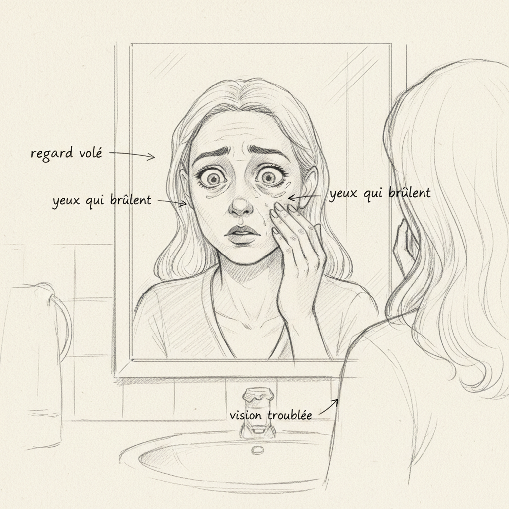
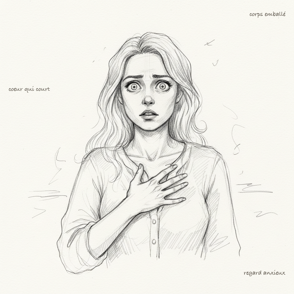
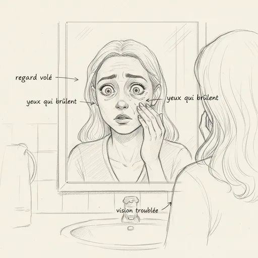
Sandrine was forty-two years old. She came to consult me six months after her Graves’ disease diagnosis, not for her thyroid, but for her eyes. Her endocrinologist had prescribed Neomercazole, her TSH was rising slowly, her palpitations had calmed down. But her eyes. Her eyes had changed. Not overnight, not dramatically. It was insidious. First a sensation of sand under her eyelids, like after a sleepless night that never ended. Then persistent redness that eye drops couldn’t soothe. And one morning, while putting on makeup, she noticed that her eyes seemed larger, more open, as if someone had pulled her eyelids upward during sleep. Her husband told her she had a fixed gaze, a look he didn’t recognize. When she showed a photo of herself from two years ago next to a recent selfie, the difference chilled her.
The ophthalmologist measured bilateral exophthalmos of twenty-two millimeters (normal is below twenty). He spoke to her of “ocular Graves’ disease,” orbitopathy, risks to vision if it progressed. He prescribed corticosteroids. No one mentioned selenium to her. No one explained what was actually happening behind her eyeballs. And no one told her that the tobacco she’d smoked for twenty years multiplied her risk of severe ocular complications sevenfold.
If you’ve read my article on Graves’ disease, you already know that exophthalmos affects about fifty percent of patients. What you may not know is that ocular involvement is often the most anxiety-provoking symptom, the most socially visible, and paradoxically the least well managed. Because endocrinologists manage the thyroid, ophthalmologists manage the eyes, and no one connects the intestine, immunity, and what’s happening behind the orbit.

What Happens Behind Your Eyes
To understand Graves’ orbitopathy, you first need to visualize the anatomy. Behind each eyeball, in the bony cavity of the orbit, lies a cushion of adipose and connective tissue that cushions the eye and allows the extraocular muscles to function properly. This tissue, normally unobtrusive, contains fibroblasts: these architect cells that maintain the structure of connective tissue. Under normal circumstances, these fibroblasts go about their business quietly, producing collagen and glycosaminoglycans in reasonable quantities.
In Graves’ disease, these fibroblasts become the site of a genuine immune invasion. CD4+ and CD8+ T lymphocytes, B lymphocytes, and macrophages massively infiltrate the retro-orbital tissue, just as they would infiltrate infected tissue. Except there’s no infection. The immune system is targeting the wrong thing, once again.
The mechanism is fascinating and revealing. Orbital fibroblasts express the TSH receptor on their surface, the same receptor as thyrocytes. And TRAb antibodies, those stimulating antibodies that rev up the thyroid, also bind to these orbital fibroblasts. When TRAb activates a thyrocyte, it produces excess thyroid hormones. When TRAb activates an orbital fibroblast, it goes haywire in another direction: it begins massively producing glycosaminoglycans, these hydrophilic molecules that attract water like a sponge. Kahaly and colleagues demonstrated this as early as 1994: the accumulation of glycosaminoglycans in retro-orbital tissue is the direct consequence of immune activation of fibroblasts.
But there’s more. Bahn and Heufelder showed in 1993 that these activated fibroblasts undergo an even more spectacular transformation: they differentiate into adipocytes, into fat cells. The retro-orbital adipose tissue literally increases in volume, as if extra fat were growing behind the eyes. This dual phenomenon, accumulation of glycosaminoglycans saturated with water plus adipogenesis, creates a mass effect that progressively pushes the eyeball forward. This is exophthalmos.
The orbit is a rigid bony cavity. It cannot expand. When the volume of contents increases, the container doesn’t move. It’s the eyeball that’s pushed forward, and this gives the prominent, fixed gaze, sometimes asymmetric, that characterizes Graves’ orbitopathy. In severe cases, the extraocular muscles themselves infiltrate and thicken, explaining diplopia, the double vision that significantly handicaps daily life.
The CAS Score: Measuring Inflammation Activity
Ophthalmologists use a standardized tool to assess orbitopathy activity: the Clinical Activity Score, or CAS. It’s a seven-point score that evaluates signs of active inflammation. Spontaneous pain at rest, pain with eye movements, eyelid redness, conjunctival redness, eyelid swelling, caruncle swelling (that small pink prominence in the inner corner of the eye), and conjunctival edema. A CAS score of three or higher out of seven indicates an active inflammatory phase, meaning the orbitopathy is progressing and it’s time to intervene.
This distinction between active and inactive phases is critical. During the active phase, which lasts an average of twelve to eighteen months, inflammation is reversible and therapeutic interventions can slow or stop progression. Intravenous corticosteroids, orbital radiotherapy, and especially the naturopathic measures I’ll detail, are most effective during this window. Once the inactive phase is reached, changes become scarring: fibrosis replaces inflammation, and only surgery can correct the consequences.
This is why time matters. Waiting, observing, “see how it evolves” without doing anything during the active phase is letting fibroblasts continue their destructive work. The recommendations of the EUGOGO group (European Group on Graves’ Orbitopathy) are clear: any moderate to severe active orbitopathy justifies treatment, and terrain management should never be postponed.
Tobacco: The Number One Enemy of Your Eyes
I’ll be direct: if you smoke and have Graves’ disease, quitting smoking is the most important thing you can do for your eyes. More important than selenium, more important than omega-3s, more important than chamomile compresses. The studies are unequivocal. Smokers have seven times the risk of developing severe orbitopathy compared to non-smokers. Seven times. This isn’t one risk factor among others, this is THE number one modifiable risk factor.
The mechanism is multifaceted. Nicotine directly stimulates orbital fibroblasts, increasing their glycosaminoglycan production and adipocyte differentiation. Carbon monoxide reduces oxygenation of orbital tissues, worsening tissue hypoxia and stimulating pathological neovascularization. Tobacco free radicals deplete selenium and vitamin E reserves, the two most protective antioxidants for orbital tissue. And cigarette smoke shifts the local immune balance in favor of pro-inflammatory cytokines.
The Bartalena study published in 1998 showed that treatment with radioactive iodine worsened orbitopathy in smokers but not in non-smokers. This result is eloquent: tobacco doesn’t just worsen existing ocular involvement, it sensitizes orbital tissues to other insults. Sandrine, my patient, smoked a pack a day for twenty years. When I showed her this data, she quit within a week. Not out of virtue, but out of fear. And sometimes fear is a more effective motivator than virtue.
The EUGOGO Study: Selenium as an Ocular Shield
In 2011, the EUGOGO study published in the New England Journal of Medicine transformed the management of mild Graves’ orbitopathy. This randomized double-blind study conducted on one hundred fifty-nine patients across several European centers compared three groups: selenomethionine at 200 micrograms daily, pentoxifylline, and placebo, for six months, with follow-up at twelve months.
The results were clear. The selenium group showed significant improvement in ocular quality of life compared to placebo. Orbitopathy progression was significantly reduced in the selenium group: only seven percent progression versus twenty-two percent in the placebo group. And clinical improvement was superior: sixty-one percent improvement in the selenium group versus thirty-six percent on placebo. Pentoxifylline, on the other hand, performed no better than placebo.
These numbers are remarkable for a simple micronutrient. How does selenium protect the eyes? Through at least three mechanisms. The first is antioxidant: selenium is the essential cofactor of glutathione peroxidase, the enzyme that neutralizes lipid peroxides generated by orbital inflammation. Activated fibroblasts produce massive quantities of free radicals, and selenium is their main antidote. The second is immunomodulatory: selenium influences T lymphocyte differentiation and reduces production of pro-inflammatory cytokines like TNF-alpha and interleukin-1. The third is direct: selenoproteins are expressed in orbital tissue and participate in local fibroblast homeostasis.
This is why selenium supplementation is recommended by the EUGOGO group in all mild Graves’ orbitopathy. Not replacing medical follow-up, but as systematic support. One hundred to two hundred micrograms of selenomethionine daily for at least six months, ideally twelve. Sunday Natural offers pharmaceutical-grade selenomethionine with ten percent discount using code FRANCOIS10.
The Naturopathic Ocular Protocol
Beyond selenium, I’ve developed a comprehensive ocular protocol in my practice for Graves’ patients. This protocol doesn’t replace ophthalmologic follow-up, it complements it. It aims to calm orbital inflammation, protect tissues against oxidative stress, and support tear film quality, which is often the first to suffer.
Vitamin E is the second antioxidant pillar after selenium. At 400 IU daily of d-alpha-tocopherol (natural form, not synthetic dl-alpha), it protects the cell membranes of orbital fibroblasts against lipid peroxidation. Selenium and vitamin E work synergistically: glutathione peroxidase (selenium-dependent) neutralizes peroxides in the aqueous phase, vitamin E neutralizes them in the lipid phase. Together, they cover both cellular compartments.
Omega-3 EPA and DHA at two to three grams daily act on systemic and local inflammation. EPA is the precursor of resolvins and protectins, anti-inflammatory molecules that accelerate resolution of inflammation rather than simply blocking it. This is an important distinction: conventional anti-inflammatories block the inflammatory process, which can delay healing. Resolvins accelerate the transition from the inflammatory phase to the repair phase. In someone whose orbital fibroblasts are in active phase, this distinction is critical.
Zinc as bisglycinate, fifteen to thirty milligrams daily, is often overlooked in the ocular context. Yet zinc is a cofactor of superoxide dismutase (SOD), the first-line antioxidant enzyme. It also participates in vitamin A metabolism and maintenance of mucosal integrity, including ocular conjunctiva. Zinc deficiency, common in autoimmune thyroid diseases, worsens both oxidative stress and vulnerability of ocular tissues.
Vitamin D deserves special attention. Several studies have shown a correlation between vitamin D deficiency and the severity of Graves’ orbitopathy. Vitamin D is a powerful immunomodulator that influences the Th1/Th2 balance and cytokine production. A 25-OH vitamin D level below 30 ng/mL justifies supplementation of 4000 to 6000 IU daily, especially since hypothyroid patients (which many Graves’ become after treatment) produce less bile and absorb fat-soluble vitamin D less well.
Local Care: Hydrate, Protect, Drain
Dry eyes are the most invalidating daily symptom in orbitopathy. When eyelids no longer close completely, especially at night, the cornea dries out. This exposure keratitis can cause corneal ulcerations if not managed.
Preservative-free artificial tears are the foundation of local treatment. Not eye drops in bottles with benzalkonium chloride, which is irritating and toxic to the corneal epithelium long-term. Preservative-free single-dose vials, used as often as needed, at least four to six times daily. At night, a more viscous ophthalmic gel protects the cornea during sleep, especially if eyelid closure is incomplete. A silk sleep mask can help maintain humidity and protect the eyes.
Cool compresses of Roman chamomile or eyebright (Euphrasia officinalis), applied to closed eyelids for ten minutes morning and evening, calm local inflammation and ease the burning sensation. Eyebright, which herbalists call “the spectacle-breaker,” has particular affinity for the ocular sphere. In concentrated infusion (two tablespoons per cup of boiling water, steeped ten minutes then cooled), it reduces eyelid edema and conjunctival redness. These aren’t placebos: eyebright contains iridoids (notably aucubin) that exert documented anti-inflammatory and astringent action on ocular mucous membranes.
Facial lymphatic drainage is an underestimated tool. Periorbital edema isn’t just inflammatory, it’s lymphatic. The accumulation of hydrophilic glycosaminoglycans in retro-orbital tissue creates liquid stasis that the lymphatic system struggles to drain. Gentle facial lymphatic drainage performed by a trained physiotherapist or osteopath, twice weekly during the active phase, can significantly reduce edema and the sensation of orbital pressure. Self-massage exercises around the orbit, with very light finger-tip pressure moving from the inner angle outward then toward the pre-auricular nodes, can be performed daily.
When the Eye Becomes an Emergency
There are signs that should never be ignored. Compression of the optic nerve by hypertrophied tissues is the most serious complication of orbitopathy. It affects about five percent of patients with severe orbitopathy, and constitutes an absolute ophthalmic emergency.
Warning signs are sudden loss of visual acuity, loss of color vision (desaturation of reds is often the first sign), sudden appearance of double vision, intense eye pain at rest that doesn’t respond to simple analgesics, and rapid swelling with intense redness of eyelids and conjunctiva. If you experience any of these signs, this isn’t the time for eyebright tea. This is the time for ophthalmic emergency care.
Surgical orbital decompression, which consists of weakening the bony walls of the orbit to create space and relieve pressure on the optic nerve, is sometimes the only option to preserve vision. It’s a major procedure, but it can save your sight. The message is clear: naturopathy accompanies, supports, modulates the terrain. It never replaces surgery when the optic nerve is at risk.
The Gut-Eyes Link: Why Seignalet Is Relevant Here Too
You might wonder what connection there is between your intestines and your eyes. The connection is the same as between your intestines and your thyroid: TRAb antibodies. As I explain in my article on the xenoimmune mechanism of Graves’, TRAb are probably xenoantibodies anti-Yersinia enterocolitica that cross-react with the TSH receptor. That same receptor is expressed by orbital fibroblasts. So reducing TRAb production by repairing leaky gut and eliminating molecular mimicry also protects the eyes.
The hypotoxic diet of Seignalet, without gluten or dairy products, with gentle cooking below 110 degrees, remains the dietary foundation. Not because it directly targets orbital fibroblasts, but because it reduces intestinal permeability, decreases passage of antigenic bacterial peptides, and lowers overall autoimmune burden. When TRAb decreases, all target organs benefit: the thyroid slows, the heart calms, and the eyes de-swell.
Individualization through IgG food analysis, as recommended by Wentz and Mouton, goes further than simply removing gluten and dairy. Each patient has their own profile of food sensitivities contributing to their intestinal permeability. A well-interpreted IgG test can reveal unsuspected reactivities, for example to eggs, soy or yeast, that silently maintain systemic and orbital inflammation.
The Importance of Stress in Ocular Involvement
Rosch’s studies show that stress is the primary trigger for Graves’ in more than ninety percent of cases. What’s less known is that stress directly influences the severity of orbitopathy. Chronically elevated cortisol shifts the local immune balance in the orbit, increasing pro-inflammatory cytokine production by infiltrating lymphocytes. Adrenaline, via beta-adrenergic receptors present on orbital fibroblasts, stimulates their proliferation and secretory activity.
This is why heart rate variability, practiced three times daily for five minutes (the 365 method: three times daily, six breaths per minute, for five minutes), isn’t optional in the ocular protocol. By activating the vagus nerve and shifting the autonomic nervous system toward the parasympathetic, it directly reduces orbital fibroblast activation. Dr Du Chazaud wrote that “the thyroid is the gland of emotion,” and this emotion is literally readable in the Graves’ patient’s eyes.
Ocular Follow-Up: What Your Ophthalmologist Should Measure
Regular ophthalmologic follow-up is essential throughout the active phase. The Hertel exophthalmometer measures eyeball protrusion in millimeters. Visual field and color vision examination detects early optic nerve involvement. Orbital MRI visualizes extraocular muscle thickening and retro-orbital tissue volume. The CAS score, re-evaluated every three months, guides therapeutic decisions.
This follow-up must be done by an ophthalmologist experienced with thyroid orbitopathy, ideally in an EUGOGO reference center. Because Graves’ orbitopathy isn’t a classical ocular pathology: it requires cross-disciplinary expertise between endocrinology, ophthalmology and immunology that not all practitioners master.
Sandrine, Six Months Later
Sandrine came back to see me after six months on protocol. She had quit smoking the very day of our first consultation. She was taking two hundred micrograms of selenomethionine daily, one thousand milligrams of omega-3 EPA/DHA, four hundred IU of natural vitamin E, and thirty milligrams of zinc bisglycinate. She was doing eyebright compresses morning and evening, heart rate variability three times daily, and had started bi-monthly facial lymphatic drainage. She’d followed the Seignalet diet since our first consultation.
Her ophthalmologist measured exophthalmos at nineteen millimeters, down from twenty-two six months earlier. Her CAS score had dropped from four out of seven to one out of seven. Dry eye had decreased markedly, and the sensation of retro-orbital pressure had nearly disappeared. Her TRAb had dropped forty percent, which explained the overall improvement in all her symptoms, including ocular.
The ophthalmologist told her he had “never seen such rapid progression.” It’s not magic. It’s applied biochemistry. When you give the body the selenium its orbital selenoproteins need, when you stop intoxicating fibroblasts with nicotine, when you reduce oxidative stress with omega-3s and vitamin E, when you calm systemic autoimmunity by repairing the gut, when you lower cortisol with heart rate variability, the eyes respond. Because the problem was never in the eyes. The problem was in the gut, in immunity, in stress, in tobacco. The eyes were just the messenger.
Want to assess your status? Take the free Claeys thyroid questionnaire in 2 minutes.
If you want personalized support, you can book a consultation.
To Go Further
- Iodine and autoimmune thyroid: danger or benefit? Separating fact from fiction
- Graves’ disease: understanding autoimmune hyperthyroidism
- Graves’: preventing relapse after Neomercazole
- Hashimoto: the forgotten causes your doctor isn’t looking for
Sources
- Marcocci, Claudio, et al. “Selenium and the Course of Mild Graves’ Orbitopathy.” New England Journal of Medicine 364.20 (2011): 1920-1931.
- Bartalena, Luigi, et al. “Relation between therapy for hyperthyroidism and the course of Graves’ ophthalmopathy.” New England Journal of Medicine 338.2 (1998): 73-78.
- Seignalet, Jean. L’Alimentation ou la Troisième Médecine. 5th ed. Paris: François-Xavier de Guibert, 2004.
- Kahaly, George J., et al. “Glycosaminoglycan synthesis in orbital connective tissue.” Thyroid 4.3 (1994): 303-308.
If you want personalized support for your Graves’ orbitopathy, you can book a consultation. I consult in my Paris office and via video throughout France. You can also contact me for any questions.
To go further, my complete thyroid training covers everything I’ve written in my thyroid articles with case studies, commented assessments and detailed protocols. And if you’re looking for the basics of naturopathy to understand terrain and detoxification pathways, it’s the best starting point.
For thyroid supplementation, Sunday Natural offers pharmaceutical-grade selenium, zinc and vitamin D (ten percent discount with code FRANCOIS10). The Inalterra grounding mat reduces nighttime autoimmune inflammation (ten percent discount with code FRANCOISB). Find all my partnerships with exclusive promo codes.
Your eyes aren’t condemned by Graves’. They’re a reflection of what’s happening in your terrain. Calm the terrain, and your eyes will calm.


Laisser un commentaire
Sois le premier à commenter cet article.