Pauline is thirty-eight years old with fatigue that hasn’t subsided for two years, hair falling out by the handful, an eight-kilogram weight gain without dietary changes, a mental fog that prevents her from concentrating at work, and low mood. She consulted three doctors. All three measured her TSH. All three found 2.8 mIU/L. All three told her that her thyroid was fine.

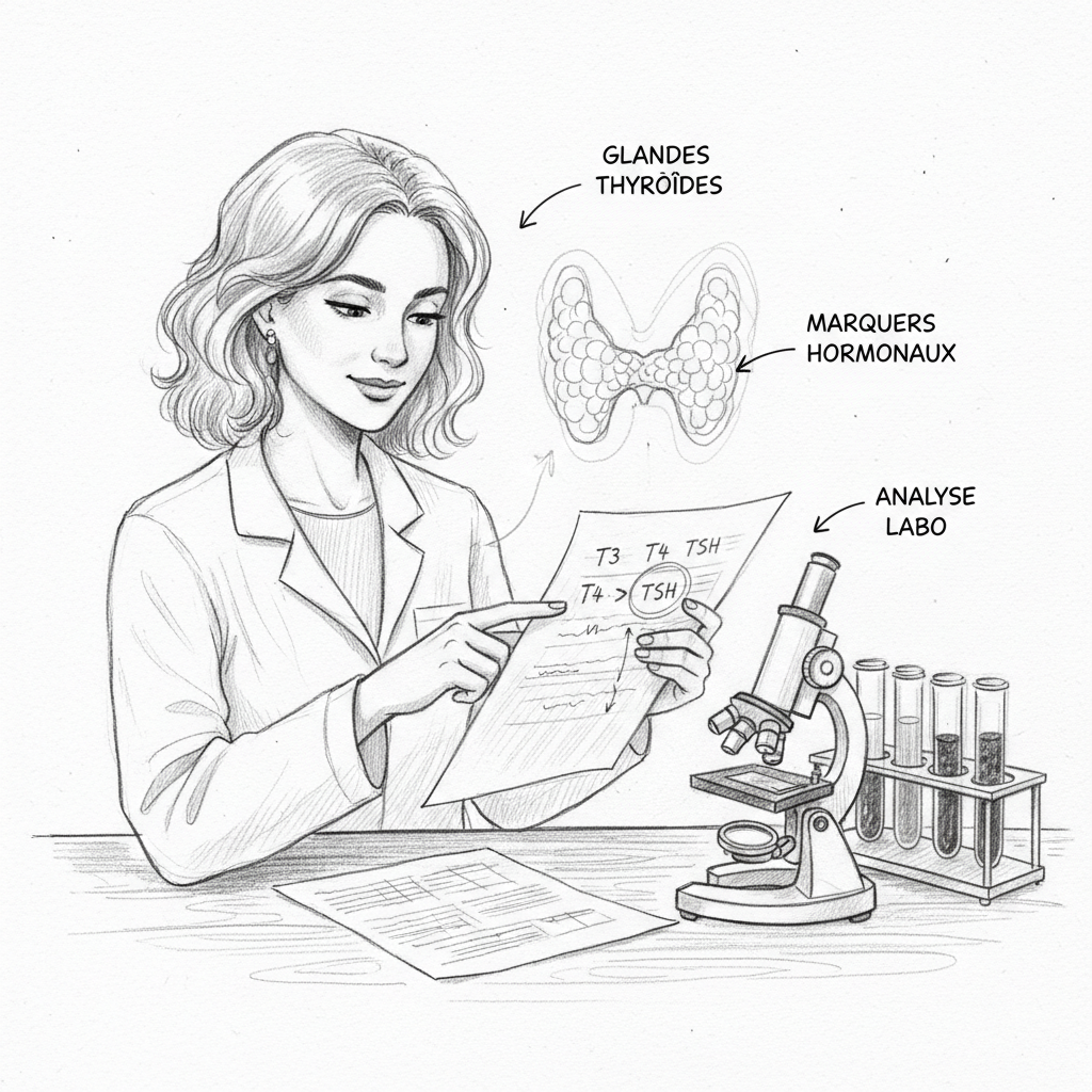
When Pauline came to my office, I requested a complete thyroid panel. Ten markers instead of just one. Here’s what we found: TSH at 2.8 mIU/L (within the “normal” range but in the upper third), free T4 at 14.2 pmol/L (correct), free T3 at 3.1 pmol/L (functionally low, a sign that the liver is not properly converting T4 to T3), anti-TPO at 180 IU/mL (positive, indicating autoimmune thyroiditis that no one had looked for), anti-thyroglobulin at 45 IU/mL (high normal), and a collapsed T3L/T4L ratio.
Pauline didn’t have “a thyroid that was fine.” Pauline had Hashimoto’s disease progressively destroying her thyroid gland, likely for years, with altered T4 to T3 conversion. All of this invisible with a simple TSH measurement.
The trap of TSH alone
TSH (Thyroid Stimulating Hormone) is the hormone secreted by the pituitary gland to stimulate the thyroid. When the thyroid produces enough hormones, TSH drops (negative feedback). When the thyroid doesn’t produce enough, TSH rises to stimulate it further. It’s an elegant mechanism that, in theory, makes TSH a faithful reflection of thyroid function.
In practice, TSH alone is a crude indicator that misses many clinical situations. The first is subclinical hypothyroidism (TSH between 2.5 and 4.5 with T3 and T4 still within normal ranges but symptoms already present). The second is impaired T4 to T3 conversion (normal TSH, normal T4, but low T3 because the liver, kidneys, or intestines aren’t converting). The third is early-stage autoimmune thyroiditis (antibodies destroy the gland for years before TSH begins to rise). The fourth is thyroid hormone resistance (hormones are produced in normal amounts but cellular receptors don’t “hear” them).
Hertoghe, in his clinical endocrinology treatise, denounces this diagnostic reduction: “Reducing thyroid assessment to TSH alone is like evaluating a company’s financial health by looking only at its revenue without examining its expenses, debts, cash flow, and profitability. Revenue can be fine while the company goes bankrupt. TSH can be normal while the patient has tissue-level hypothyroidism.”
The ten markers of the complete panel
The first marker is TSH. It’s the starting point, not the destination. The standard laboratory norm is 0.4 to 4.0 mIU/L (or even 4.5 according to some labs). The optimal functional norm falls between 0.5 and 2.0 mIU/L. Above 2.5, especially with symptoms, further investigation is needed. Below 0.5, hyperthyroidism or Levothyroxine overdose must be excluded.
The second marker is free T4 (levothyroxine). It’s the storage hormone produced by the thyroid. The laboratory norm is 9 to 19 pmol/L. The functional norm is in the upper third, between 14 and 18 pmol/L. Low free T4 with normal TSH can indicate central hypothyroidism (pituitary issue) or iodine deficiency.
The third marker is free T3 (triiodothyronine). It’s the active hormone, the one that enters cells and accelerates metabolism. Only 20% of T3 is produced directly by the thyroid; the remaining 80% comes from T4 to T3 conversion in the liver, kidneys, and intestines. The functional norm falls in the upper third of the reference range. It’s THE marker that doctors measure least and yet is most strongly correlated with clinical symptoms.
The fourth marker is the T3L/T4L ratio. This ratio assesses the efficiency of peripheral conversion. A low ratio (correct T4 but low T3) indicates a conversion problem, often linked to hepatic overload, selenium or zinc deficiency, chronic stress (cortisol inhibits deiodinase), or systemic inflammation.
The fifth marker is reverse T3 (rT3). When the body is stressed, inflamed, or in caloric restriction, it preferentially converts T4 to inactive reverse T3 rather than active T3. This is an energy-saving mechanism that becomes pathological when it persists. High rT3 with low T3 is the classic picture of “low T3 syndrome” seen in chronic stress, serious illness, restrictive diets, and adrenal fatigue.
The sixth marker is anti-TPO antibodies (anti-thyroperoxidase). TPO is the key enzyme in thyroid hormone synthesis. Positive anti-TPO antibodies (above 35 IU/mL) indicate autoimmune thyroiditis (Hashimoto’s). It’s the most sensitive autoimmune marker. It can be positive ten to fifteen years before TSH begins to rise, making it an invaluable early detection tool.
The seventh marker is anti-thyroglobulin antibodies (anti-TG). Thyroglobulin is the precursor protein for thyroid hormones. Anti-TG is positive in approximately 80% of Hashimoto’s patients, often in association with anti-TPO. But in 10 to 15% of patients, anti-TG is the only positive antibody, highlighting the importance of measuring both.
The eighth marker is thyroglobulin. It’s mainly used in post-cancer thyroid follow-up, but it can also reflect active thyroid inflammation in Hashimoto’s (elevated thyroglobulin: tissue destruction in progress).
The ninth marker is urinary iodine. Iodine is the fundamental substrate for hormone synthesis (four iodine atoms are needed to make a T4 and three for a T3). 24-hour urine iodine evaluates iodine status. In France, 30 to 40% of the population has moderate iodine insufficiency, which silently contributes to thyroid dysfunction. But beware: iodine supplementation is a delicate matter in Hashimoto’s patients because excess iodine can aggravate autoimmunity.
The tenth marker is thyroid ultrasound. It’s not a blood marker but an indispensable complement to the panel. Ultrasound reveals gland size, vascularization, texture (homogeneous or heterogeneous), presence of nodules, and signs of thyroiditis (hypoechoic, heterogeneous, pseudonodular appearance). In patients with negative antibodies but suggestive symptoms, ultrasound can confirm seronegative Hashimoto’s.
Functional norms versus laboratory norms
This might be the most important point in this article. Laboratory norms (also called reference intervals) are calculated statistically from a “reference” population sample. The problem is that this sample often includes undiagnosed people who themselves have thyroid dysfunction. Result: the norms are artificially wide.
The most blatant example is TSH. The standard norm of 0.4 to 4.0 mIU/L means a patient at 3.8 is considered “normal.” Yet epidemiological studies show that 95% of healthy people (without thyroid pathology) have TSH below 2.5 mIU/L. The AACE (American Association of Clinical Endocrinologists) even proposed in 2003 to tighten the upper norm to 3.0 mIU/L, then to 2.5 mIU/L, a proposal never universally adopted but reflecting clinical reality: many patients “normal” on paper are hypothyroid in practice.
Curtay, in his nutritherapy approach, distinguishes “statistical norms” (which say “you’re not sick”) from “optimal norms” (which say “your body is functioning optimally”). This distinction is fundamental in functional naturopathy. A patient with TSH at 3.5, free T3 in the lower third, and hypothyroid symptoms is not “normal.” He or she is in a gray zone that conventional medicine doesn’t yet recognize but that functional medicine has been treating for twenty years.
Seronegative Hashimoto’s
One particular case deserves attention. Approximately 10 to 15% of patients with histologically confirmed Hashimoto’s thyroiditis (by biopsy or surgery) never have detectable antibodies in their blood. This is seronegative Hashimoto’s.
Antibodies are present in thyroid tissue but don’t (or barely) enter the bloodstream, likely because the autoimmune attack is localized and immune complexes are trapped in the gland. Ultrasound then becomes the key diagnostic tool: it shows a typically Hashimoto appearance (hypoechoic, heterogeneous, pseudonodular) in the absence of circulating antibodies.
This is why I routinely recommend thyroid ultrasound to complement the blood panel, especially when clinical symptoms are suggestive but antibodies come back negative.
How to obtain this panel in France
In France, only TSH, free T4, and anti-TPO antibodies are reimbursed as first-line tests (and even then, anti-TPO is only reimbursed if TSH is abnormal, which is a vicious circle). Free T3, anti-TG, rT3, and urinary iodine are generally not prescribed routinely and may require a specific order.
A few practical tips: ask your doctor for an order including TSH, T4L, T3L, anti-TPO, and anti-TG by explaining your symptoms. If your doctor refuses T3L and antibodies, some labs accept “off-nomenclature” testing (not reimbursed, between 15 and 30 euros per marker). rT3 is rarely available in French community labs and may require sending to a specialized laboratory. Urinary iodine is done on 24-hour urine and requires specific collection.
Blood should be drawn in the morning before 9am, fasting, without having taken Levothyroxine (take it after). If you’re taking biotin (often present in “hair and nails” supplements), stop it at least 48 hours before because it interferes with immunological assays and can artificially falsify TSH downward and T4 upward, mimicking hyperthyroidism that doesn’t exist.
Interpreting the panel: patterns to know
The classic pattern of frank hypothyroidism: elevated TSH (above 4.5), low T4L, low T3L. This is the easy case, diagnosed and treated by all doctors.
The pattern of subclinical hypothyroidism: TSH between 2.5 and 4.5, T4L in the lower half, T3L in the lower third. Symptoms are present but the doctor says “everything is normal.” This is the most frustrating and most frequent case in my practice.
The pattern of conversion defect: normal or slightly elevated TSH, correct T4L (upper third), low T3L (lower third). The liver isn’t converting. This pattern is common in patients with hepatic overload, SIBO, chronic inflammation, or prolonged stress. Hepatic support (milk thistle, castor oil compress) and correction of cofactors (selenium, zinc, iron) are the therapeutic levers.
The pattern of thyroid hormone resistance: normal TSH, normal T4L and T3L, but frank hypothyroid symptoms. Hormones are produced but cells don’t “hear” them. This pattern is often linked to systemic inflammation (which reduces thyroid receptor expression), insulin resistance, or heavy metal toxicity.
Warning
The complete thyroid panel is a tool for understanding, not self-diagnosis. Fine interpretation of results, correlation with clinical presentation, and therapeutic decisions (especially introduction or adjustment of Levothyroxine) are the physician’s responsibility. The naturopath provides a complementary functional perspective and natural support tools (micronutrition, diet, stress management), but does not replace medical follow-up.
Masson, in his practice of experiential dietetics, reminded us that “biology is just a snapshot at one moment in time. The clinician reads the story, biology confirms it.” This is why the complete thyroid panel doesn’t replace listening to the patient. Pauline had clear symptoms for two years. Her body was speaking. It just took listening and ordering the right tests to confirm what her body was already saying.
Want to evaluate your status? Take the free Claeys thyroid questionnaire in 2 minutes.
If you want personalized support, you can book a consultation.

Laisser un commentaire
Sois le premier à commenter cet article.