Her name was Monique (name changed), 62 years old, a retired teacher. When she sat down across from me, she had a medical file under her arm and that resigned expression I recognize in patients who bounce from specialist to specialist without ever getting a comprehensive answer. “I was diagnosed with lumbar osteoporosis two years ago. My cardiologist found calcifications on my coronary arteries. And my doctor tells me it’s just age.” Monique had been taking a proton pump inhibitor for eight years for acid reflux. Vitamin D for three years, prescribed by her rheumatologist. And calcium, on her pharmacist’s advice. No one had ever mentioned vitamin K to her.
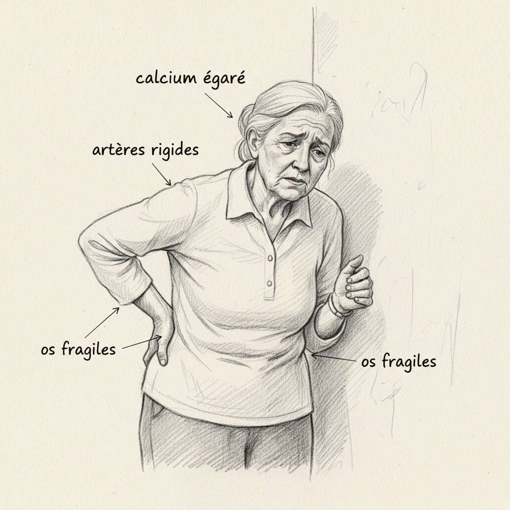
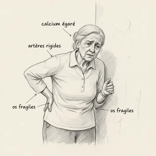
When I looked at her file, the paradox jumped out at me. The calcium she swallowed every morning wasn’t binding to her bones. It was depositing in her arteries. Her body was losing calcium where it needed it, and accumulating it where it shouldn’t. This phenomenon has a name in biochemistry: the calcium paradox. And the key to this paradox is a vitamin that almost nobody talks about, that almost nobody tests for, and that almost nobody supplements correctly: vitamin K.
“You can’t understand osteoporosis without understanding vitamin K. And you can’t understand arterial calcifications without understanding vitamin K. It’s the missing link between bones and arteries.” Jean-Paul Curtay
Vitamin K is the great forgotten nutrient of micronutrition. Discovered in 1929 by Danish biochemist Henrik Dam, who received the Nobel Prize in 1943 for this work, it gets the K from “Koagulation” in Danish. For decades, it was reduced to this single role: blood coagulation. Then research revealed something much vaster. Vitamin K doesn’t just make your blood clot. It directs calcium in your body. It sends it to your bones, and it prevents it from depositing in your arteries. Without it, calcium becomes a missile without guidance. It binds in the wrong place. And the consequences are devastating.

The causes of vitamin K deficiency
To understand the deficiency, you first need to understand the two forms of this vitamin, because they have neither the same sources, nor the same roles, nor the same metabolism. Vitamin K1 (phylloquinone) is synthesized by green plants. It participates in photosynthesis and concentrates in the leaves. When you eat spinach, kale, broccoli, parsley, you ingest K1. This K1 is captured primarily by the liver, where it serves as a cofactor to gamma-glutamyl carboxylase, the enzyme that activates coagulation factors II (prothrombin), VII, IX and X. Without K1, these factors remain undercarboxylated, inactive, and blood doesn’t clot properly. This is why the newborn, whose intestinal microbiota is still sterile and whose hepatic vitamin K reserves are nearly nonexistent, systematically receives a vitamin K injection at birth to prevent hemorrhagic disease of the newborn1.
Vitamin K2 (menaquinone) is a completely different story. It exists in several forms, designated MK-n according to the length of their side chain. The two most studied forms are MK-4 and MK-7. MK-4 has a short half-life, on the order of two hours, which means it disappears quickly from the blood. MK-7, on the other hand, has a half-life of 72 hours2. Three days. This long persistence in the blood allows it to reach extra-hepatic tissues that K1 cannot reach: bones, arteries, teeth, cartilage. And that’s where everything happens.
K2 comes from two sources. The first is bacterial fermentation. The bacteria in your intestinal microbiota, particularly Bacteroides fragilis, Escherichia coli and certain strains of Lactobacillus, synthesize K2. This isn’t trivial: in a healthy gut, this endogenous production covers a significant portion of needs. The second source is fermented foods: Japanese natto (soy fermented by Bacillus subtilis), aged hard cheeses, sauerkraut, miso. The MK-7 K2 from natto is the most studied form in clinical research. It’s also the form that circulates best in the body.
The first cause of deficiency is therefore a diet poor in green vegetables (K1 deficit) and devoid of fermented foods (K2 deficit). Modern Western diet checks both boxes. Industrial meals contain few green leaves and no traditional fermented foods. The second cause is intestinal dysbiosis. When the microbiota is imbalanced by antibiotics, stress, a diet rich in refined sugars or chronic candidiasis, the bacteria that produce K2 are decimated. Endogenous production collapses. The third cause is fat malabsorption. Vitamin K is fat-soluble, which means it needs dietary fats and bile salts to be absorbed in the small intestine. Any condition that disrupts fat digestion (bile insufficiency, cholestasis, celiac disease, Crohn’s disease, ileal resection, cystic fibrosis) compromises vitamin K absorption. It’s the same mechanism as for vitamin D: no fat, no absorption.
The fourth cause is iatrogenic, and it’s massive. Proton pump inhibitors (PPIs), prescribed in millions in France for acid reflux, reduce gastric acidity and disrupt the absorption of all fat-soluble vitamins, including K. AVK-type anticoagulants (warfarin, fluindione, acenocoumarol) deliberately block vitamin K recycling to thin the blood. And broad-spectrum antibiotics destroy the bacteria that produce K2. When a patient combines long-term PPIs, repeated antibiotic therapy and a diet poor in green vegetables, the conditions for deficiency are met. This was exactly Monique’s situation.
The symptoms of deficiency
Vitamin K deficiency doesn’t manifest as a single sign, but as a constellation of problems affecting three distinct systems: coagulation, bones and arteries. This dissociation is what makes diagnosis so difficult, because doctors monitoring coagulation don’t look at bones, those monitoring bones don’t look at arteries, and nobody connects it to vitamin K.
The first axis is coagulation. K1 activates coagulation factors II, VII, IX and X, as well as anticoagulant proteins C and S. In severe deficiency, prothrombin time (PT) lengthens and INR increases. Clinical signs include easy bruising (bruises from minor bumps), gum bleeding, nosebleeds, heavy menstrual bleeding. In infants, hemorrhagic disease can cause fatal intracranial hemorrhages, which justifies systematic K1 injection at birth. In routine practice, isolated K1 deficiency is rare in adults whose diet contains a minimum of green vegetables. The problem lies elsewhere.
The second axis is bones. And that’s where K2 enters the scene. Osteocalcin is a protein synthesized by osteoblasts (cells that build bone). To be functional, it must be carboxylated by gamma-glutamyl carboxylase, a K-dependent enzyme. Carboxylated osteocalcin binds calcium in the bone matrix. It’s literally the cement that integrates calcium into bone hydroxyapatite. Without vitamin K, osteocalcin remains undercarboxylated, inactive. The calcium you absorb thanks to vitamin D and your diet doesn’t bind to your bones. It floats in the blood. And it deposits elsewhere.
The study by Knapen et al. (2015), published in Osteoporosis International, demonstrated that supplementation with 180 mcg of K2 MK-7 for three years significantly reduced bone mineral density loss and improved mechanical resistance of vertebrae in postmenopausal women3. Three years of follow-up, 244 women, measurable results. This is proof that K2 doesn’t just slow bone loss: it actively contributes to bone strength. Yet how many rheumatologists prescribe it? Almost none. We prescribe calcium and vitamin D. We forget K2. And we’re amazed that osteoporosis progresses.
The third axis is arteries. MGP (Matrix Gla Protein) is a protein secreted by smooth muscle cells in arterial walls and chondrocytes in cartilage. Like osteocalcin, it must be carboxylated by a K-dependent enzyme to be active. Carboxylated MGP is the most powerful natural inhibitor of soft tissue calcification known to date. It prevents calcium from depositing in arterial walls, heart valves, kidneys. Without vitamin K, MGP remains inactive, and arteries calcify progressively. Walls become rigid, blood pressure rises, cardiovascular risk skyrockets.
The Rotterdam Study, conducted by Geleijnse et al. on 4807 subjects followed for ten years, is one of the most impressive in all nutritional literature4. The results are unambiguous: participants with the highest intakes of vitamin K2 had a 57% reduced risk of cardiovascular mortality and a 52% reduced risk of aortic calcification compared to those with the lowest intakes. And the crucial point: vitamin K1 had no protective effect on arteries. Only K2 mattered. This confirms that K1 remains captured by the liver for coagulation, while K2 reaches peripheral tissues (arteries, bones) where it activates MGP and osteocalcin.
The calcium paradox is therefore the following: a patient deficient in K2 loses bone calcium (osteoporosis) while accumulating arterial calcium (calcified atherosclerosis). Bones empty out, arteries stiffen. This was exactly what was happening to Monique. And it was exactly what nobody had explained to her.

The essential micronutrients for vitamin K
Vitamin K doesn’t function alone. It exists within a network of biochemical synergies whose three pillars are vitamin D, magnesium and dietary calcium. Understanding these interactions means understanding why supplementing a single nutrient without the others is not only ineffective, but potentially dangerous.
Vitamin D increases intestinal calcium absorption (via the calbindin transport protein) and stimulates osteocalcin synthesis by osteoblasts. The more active vitamin D you have, the more osteocalcin your osteoblasts produce. But this osteocalcin is secreted in inactive form (undercarboxylated). It’s vitamin K2 that carboxylates it and makes it functional. Taking vitamin D without K2 is like manufacturing locks without forging keys. You absorb more calcium, your bones produce more osteocalcin, but nothing assembles. Calcium circulates in excess in the blood, MGP remains inactive, and arteries calcify. This is the exact mechanism observed in patients supplemented with high-dose D3 without K2: poor improvement in bone density, and sometimes worsening of vascular calcifications.
Dr. Thierry Hertoghe, in his practice as an endocrinologist specialized in hormonology, systematically prescribes the combination D3 + K2 MK-7. He considers this association as a non-negotiable standard of anti-aging medicine. Jean-Paul Curtay, in his Nutritherapy, classifies K2 among “second-line” nutrients that should be supplemented in all subjects at cardiovascular and bone risk. In naturopathy, I follow this same logic: never D3 without K2. It’s a basic protocol.
Magnesium works at two levels. First, it’s a cofactor for hepatic and renal hydroxylases that activate vitamin D. Without magnesium, vitamin D remains in inactive form, calcium absorption drops, and the K-dependent cascade cannot function properly. Second, magnesium directly participates in bone mineralization: approximately 60% of body magnesium is found in bones, integrated into the hydroxyapatite matrix. A magnesium deficit weakens bone independent of vitamin K, and correcting both together is much more effective than correcting one without the other. This is the concept of the “bone triad” I use in consultation: D3 + K2 + magnesium. Everything else is secondary.
Zinc plays an auxiliary but non-negligible role. It’s a cofactor for bone alkaline phosphatase, an enzyme essential for mineralization. Postmenopausal women deficient in zinc show accelerated bone loss that only partially responds to calcium and D3 supplementation. Adding zinc to the protocol improves results measurably.
Food sources
The sources of K1 and K2 are radically different, and this distinction explains why you can very well eat five servings of vegetables a day and still be deficient in K2.
K1 is found in leafy green vegetables. Fresh parsley is the most concentrated source, with approximately 1640 mcg per 100 grams. Kale provides 817 mcg/100g, cooked spinach 494 mcg/100g, cooked broccoli 141 mcg/100g, romaine lettuce 174 mcg/100g, Brussels sprouts 177 mcg/100g. Vegetable oils (rapeseed, soy, olive) contain it too, but in more modest amounts. K1 is relatively heat-resistant, but gentle cooking remains preferable to preserve overall cofactors. The essential point: absorption of K1 from vegetables is low, on the order of 5 to 15%, because it’s trapped within the chloroplasts of plant cells. Chopping vegetables, steaming them gently and consuming them with a fat source (olive oil, butter) considerably improves this absorption.
K2 is found in fermented foods and animal products. The absolute champion is natto, this traditional Japanese fermented soy, which contains approximately 1000 mcg of K2 MK-7 per 100 grams. This is an extraordinary concentration, and it’s one of the reasons why Japanese people who regularly consume natto have osteoporotic fracture rates much lower than Westerners, despite significantly lower calcium intakes5. Aged gouda provides approximately 75 mcg/100g of K2, brie about 50 mcg/100g, emmental 43 mcg/100g. Sauerkraut provides 4.8 mcg/100g. Poultry liver (chicken, duck, goose) contains between 12 and 14 mcg/100g in MK-4 form. Egg yolks from free-range chickens provide 15 to 30 mcg/100g depending on the chickens’ diet. Butter from grass-fed cows contains significantly more K2 than industrial butter.
What these figures show is that the standard Western diet is structurally deficient in K2. Outside of natto (which almost nobody eats in France), food sources of K2 are modest and insufficient to cover optimal needs estimated at between 100 and 200 mcg per day. This is why K2 MK-7 supplementation is so relevant, especially in postmenopausal women, elderly people and patients on AVK or PPIs.
The antagonists of vitamin K
Certain factors actively destroy, block or deplete your vitamin K reserves. Knowing them is the first step to stopping the drain before refilling the tank.
AVK anticoagulants (warfarin, fluindione, acenocoumarol) are the most powerful and most commonly used antagonists. Their mechanism of action is clear: they block the VKOR enzyme (vitamin K epoxide reductase), which recycles vitamin K after each carboxylation reaction. Normally, vitamin K functions in a cycle: it’s oxidized by gamma-glutamyl carboxylase when it activates coagulation factors, then regenerated by VKOR to serve again. AVKs break this cycle. Oxidized vitamin K is no longer recycled, coagulation factors remain inactive, and blood becomes thinner. This is the desired effect in patients at thrombotic risk. But this blockade of the vitamin K cycle also affects osteocalcin and MGP. This is the AVK paradox: in thinning the blood, they simultaneously accelerate osteoporosis and arterial calcification. Studies show that patients on long-term warfarin have significantly lower bone mineral density and higher coronary calcification scores than controls6. This is a side effect rarely mentioned in the package insert, and it’s a silent tragedy for millions of treated patients.
Broad-spectrum antibiotics constitute the second major antagonist. Fluoroquinolones (ciprofloxacin, levofloxacin), third-generation cephalosporins and carbapenems decimate intestinal bacteria that produce K2. After ten days of fluoroquinolone treatment, endogenous K2 production can drop dramatically and take weeks to recover, the time it takes for the microbiota to regenerate. Risk is maximal when a patient simultaneously takes an AVK and an antibiotic: the antibiotic reduces intestinal K2 production, which amplifies the anticoagulant effect of the AVK and can cause severe hemorrhages. This interaction is documented, and it should systematically result in enhanced INR monitoring during and after antibiotic therapy.
Cholestyramine and orlistat are two medications that interfere with fat absorption, and therefore with absorption of all fat-soluble vitamins (A, D, E, K). Cholestyramine, prescribed to lower cholesterol, sequesters bile acids in the digestive tract. Without bile, no fat digestion, and without fat digestion, no vitamin K absorption. Orlistat, an anti-obesity medication, inhibits pancreatic lipases and reduces fat absorption by 30%. Patients on these treatments should systematically receive fat-soluble vitamin supplementation, yet this rarely happens.
PPIs (omeprazole, esomeprazole, pantoprazole) constitute an indirect but formidable antagonist. By reducing gastric acidity, they disrupt overall digestion, modify intestinal microbiota (favoring bacteria that don’t produce K2) and reduce absorption of fat-soluble nutrients. Long-term PPI use is associated with increased risk of osteoporotic fracture, a link partly explained by depletion in vitamin K and magnesium. Monique had been taking PPIs for eight years. Eight years of silent sabotage of her vitamin K, magnesium and calcium.
Pathological fat malabsorption (celiac disease, Crohn’s disease, pancreatic insufficiency, cholestasis) is an organic cause of deficiency. Any disease affecting the bile or proximal small intestine mucosa can compromise vitamin K absorption. And chronic alcoholism, by damaging both the liver and microbiota, combines multiple depletion factors.
The forgotten causes of deficiency
Beyond the classic causes, there are mechanisms of deficiency that conventional medicine systematically ignores, but that naturopathy, through its approach to terrain, knows how to identify.
The first forgotten cause is chronic dysbiosis without antibiotic therapy. You might not have taken any antibiotics in years and still have a devastated microbiota. Chronic stress, which modifies bacterial profiles via the gut-brain axis. A diet rich in refined sugars and ultra-processed foods, which feeds fermentable bacteria at the expense of K2-producing strains. Dietary endocrine disruptors, which modify the microbiome in still poorly understood ways. Glyphosate, classified as a broad-spectrum bacterial antibiotic in a 2010 patent, which decimates soil bacteria and potentially ours via food residues. A patient consuming non-organic bread, industrial dairy and treated fruits accumulates glyphosate traces that progressively weaken their K2-producing bacteria.
The second forgotten cause is bile salt deficiency. Bile isn’t only necessary for digesting fats. It’s essential for forming micelles that transport fat-soluble vitamins across the intestinal mucosa. Patients whose gallbladder has been removed (cholecystectomy), patients with hepatic steatosis (non-alcoholic fatty liver, which now affects 25% of the Western population), and patients whose liver is clogged by years of metabolic toxemia produce less bile and absorb vitamin K less effectively. This is a silent cause I frequently encounter in consultation, especially in postmenopausal women who combine hepatic overload, PPIs and a diet poor in green vegetables.
The third forgotten cause is high-dose vitamin E supplementation. Vitamin E antagonizes vitamin K at high doses, probably through competition for gamma-glutamyl carboxylase. Studies have shown that supplementation with 1000 IU of vitamin E daily increased coagulation time and undercarboxylated osteocalcin markers. This phenomenon is particularly relevant in patients simultaneously taking vitamin E “for heart health” and warfarin for coagulation, a combination that potentiates hemorrhagic risk.
The fourth forgotten cause is age. Aging reduces microbiota diversity (fewer K2-producing bacteria), decreases bile secretion (less absorption of fat-soluble K), reduces hepatic storage capacity and slows VKOR recycling. People over 70 combine all these factors. And it’s precisely this population that most needs K2 to protect their bones and arteries. Dr. Hertoghe considers K2 MK-7 supplementation as a first-line anti-aging prevention measure, on par with vitamin D and magnesium.
The fifth forgotten cause is menopause itself. The drop in estrogen accelerates bone remodeling by unbalancing the osteoclast/osteoblast balance in favor of resorption. Osteoclasts (cells that resorb bone) become hyperactive, while osteoblasts (cells that build bone) slow down. This imbalance increases vitamin K2 needs to activate osteocalcin produced by remaining osteoblasts. Women in perimenopause and post-menopause have significantly higher rates of undercarboxylated osteocalcin than premenopausal women, testifying to a K2 functional deficit worsened by hormonal transition.
Dietary supplements
Vitamin K2 supplementation is one of the most documented and safest in all of micronutrition. No toxicity has been reported even at high doses, making it a nutrient with a very wide therapeutic margin.
The form I recommend first-line is K2 MK-7. Its 72-hour half-life allows it to reach stable plasma concentration with a single daily dose. It effectively carboxylates osteocalcin and MGP in extra-hepatic tissues. The Knapen study (2015) used 180 mcg/day of MK-7 for three years, with significant results on bone density and vertebral resistance3. This is the reference dose. In naturopathic practice, the common dosing range is between 100 and 200 mcg per day of K2 MK-7. In high-risk patients (manifest osteoporosis, arterial calcifications, long-term AVK, menopause with low T-score), one can increase to 300 mcg/day under supervision.
K2 MK-4, unlike MK-7, was studied in Japan at pharmacological doses of 45 mg per day (yes, milligrams, not micrograms) for osteoporosis treatment. At these doses, it showed significant reduction in vertebral fractures. But its two-hour half-life requires multiple daily doses, making compliance difficult. In routine supplementation, MK-7 is much more practical and more effective at nutritional dose.
The D3 + K2 association is the basic protocol. I never prescribe vitamin D3 alone. This is a rule I set for myself once I understood the mechanism of the calcium paradox. D3 opens the calcium tap (increased intestinal absorption, osteocalcin synthesis stimulated). K2 directs the flow (activation of osteocalcin to bind calcium to bones, activation of MGP to prevent deposit in arteries). Opening the tap without directing the water is flooding the house. Dr. Curtay is categorical: “Every prescription of vitamin D should be accompanied by vitamin K2.” Magnesium completes the trio, because it’s a cofactor for vitamin D activation and because 60% of body magnesium is found in bones.
The protocol I use in consultation for postmenopausal women with osteopenia or osteoporosis:
- Vitamin D3: 2000 to 4000 IU/day, with the fattiest meal
- Vitamin K2 MK-7: 100 to 200 mcg/day, with the same meal
- Magnesium bisglycinate: 300 to 400 mg/day, morning and evening
- Zinc bisglycinate: 15 to 25 mg/day, evening
This protocol is adjusted according to biological testing: 25-OH-D3 level (objective 50-80 ng/mL), undercarboxylated osteocalcin if available, follow-up bone densitometry at twelve months. And it’s always accompanied by work on digestive terrain, because supplementing without absorbing is throwing money out the window.
An absolute point of vigilance: patients on AVK should not modify their vitamin K intake without discussing it with their doctor or cardiologist. Vitamin K opposes the action of AVKs, and abrupt supplementation can destabilize INR and provoke thrombotic risk. This doesn’t mean AVK patients are condemned to K2 deficiency. Some specialized protocols use low, stable doses of K2 MK-7 (45 mcg/day) while adjusting AVK dose in parallel, thus stabilizing INR while protecting bones and arteries. But this requires close medical monitoring and is not self-medication.
Want to assess your vitamin K deficiency risk? Take the vitamin K deficiency questionnaire in 2 minutes. And if you also suspect a problem with vitamin D or magnesium, dedicated questionnaires complete the assessment.
Back to Monique. Once I understood her picture, the strategy became clear. First, work with her doctor to reassess the relevance of PPIs after eight years of use. In many cases, reflux can be managed through lifestyle measures (elevating the head of the bed, eating earlier in the evening, avoiding acidic foods at dinner) and bitter plants that support gastric motility. Second, introduce K2 MK-7 at 200 mcg per day, combined with the D3 she was already taking. Third, stop supplemental calcium (which without K2 was fueling her arterial calcifications) and replace it with food calcium (sardines with bones, aged hard cheeses, mineral water), safer and better integrated. Fourth, add magnesium bisglycinate at 400 mg/day to support D3 activation and bone mineralization. And fifth, restore her microbiota with an intestinal rebalancing protocol to restart endogenous K2 production.
Six months later, Monique sent me a message. Her bone densitometry showed stabilization of her lumbar T-score. Her cardiologist noted that her coronary calcifications hadn’t progressed, which was already a result. And most importantly, she’d been able to cut her PPIs in half with her doctor’s agreement. She didn’t feel cured. She felt understood. And that’s often it, the real gift of naturopathy: not a magic pill, but an explanation that makes sense.
For K2 MK-7 supplementation, Sunday Natural offers K2 MK-7 extracted from natto (all-trans, the only biologically active form), combined with D3 and pharmaceutical-grade magnesium bisglycinate (-10% with code FRANCOIS10). Find all my partnerships with exclusive promo codes.
To go further
- Acetylcholine: the forgotten neurotransmitter of your memory
- Oxidative balance: Dr. Brack’s test to measure your oxidative stress
- Carnitine and thyroid: the molecule nobody tests for
- Dopamine: when motivation fades without reason
Sources
- Curtay, Jean-Paul. Nutritherapy. Marco Pietteur, 2016.
- Hertoghe, Thierry. The Hormone Handbook. 2nd ed. Luxembourg: International Medical Books, 2012.
- Mouton, Georges. Digestive Ecology. Marco Pietteur, 2004.
- Vermeer, Cees. “Vitamin K: The Effect on Health beyond Coagulation.” Frontiers in Nutrition 3 (2012): 1-8.
“Don’t kill the mosquitoes, drain the swamp. When the terrain is healthy, symptoms disappear on their own.” Pierre-Valentin Marchesseau
Scientific references
Footnotes
-
Shearer, Martin J. “Vitamin K Deficiency Bleeding (VKDB) in Early Infancy.” Blood Reviews 23, no. 2 (2009): 49-59. PMID: 18804903. ↩
-
Schurgers, Leon J., and Cees Vermeer. “Determination of Phylloquinone and Menaquinones in Food. Effect of Food Matrix on Circulating Vitamin K Concentrations.” Haemostasis 30, no. 6 (2000): 298-307. PMID: 11356998. ↩
-
Knapen, Marjo H.J., et al. “Three-Year Low-Dose Menaquinone-7 Supplementation Helps Decrease Bone Loss in Healthy Postmenopausal Women.” Osteoporosis International 24, no. 9 (2013): 2499-2507. PMID: 23525894. ↩ ↩2
-
Geleijnse, Johanna M., et al. “Dietary Intake of Menaquinone Is Associated with a Reduced Risk of Coronary Heart Disease: The Rotterdam Study.” Journal of Nutrition 134, no. 11 (2004): 3100-3105. PMID: 15514282. ↩
-
Kaneki, Masataka, et al. “Japanese Fermented Soybean Food as the Major Determinant of the Large Geographic Difference in Circulating Levels of Vitamin K2: Possible Implications for Hip-Fracture Risk.” Nutrition 17, no. 4 (2001): 315-321. PMID: 11369171. ↩
-
Caluwe, Liesbeth, et al. “Vitamin K2 Supplementation in Haemodialysis Patients: A Randomized Dose-Finding Study.” Nephrology Dialysis Transplantation 29, no. 7 (2014): 1385-1390. PMID: 24285428. ↩


Laisser un commentaire
Sois le premier à commenter cet article.