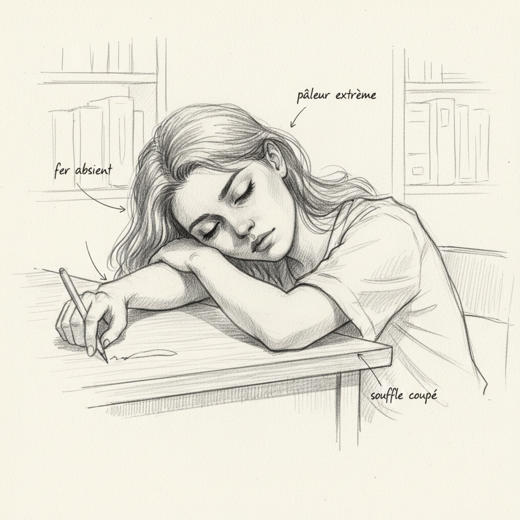
Her name is Claire (name changed), 36 years old, two children. When she sat down across from me, she had dark circles under her eyes, pale lips, and that weariness in her voice that I now recognize from three meters away. “I’ve been tired for two years. My doctor told me my blood work was normal.” I asked to see her results. Hemoglobin at 12.1 g/dL, within normal range. Ferritin at 14 ng/mL. Within laboratory “normal” range, yes. Within the normal range of health, certainly not. Claire had been iron deficient for probably years, and no one had told her.
“The good doctor is one who knows how to be a minister of vital force.” Paul Carton
Anemia is the most widespread nutritional disorder on the planet. A quarter of the world’s population is affected, meaning nearly two billion human beings[^1]. And yet, conventional medicine often treats it in a cursory manner: one iron tablet, a prescription, goodbye. No one wonders why iron isn’t being retained. No one looks at the stomach, intestines, liver, or inflammation. No one seeks the cause of the cause.
In five years of consultations, I’ve lost count of the number of patients who told me the same thing: “I took iron for six months and my ferritin didn’t budge.” That’s normal. Giving iron to a body that can’t absorb it is like watering a plant in a pot with a hole in it. As long as you don’t plug the hole, the water runs right through. And the hole, in naturopathy, we know where to look for it. That’s exactly what I’m going to detail for you in this article.
Anemia: much more than just an iron deficiency
Let’s start with the basics. Anemia, strictly speaking, is a decrease in hemoglobin levels in the blood. Hemoglobin is that protein present in red blood cells that binds to oxygen and transports it from the lungs to every cell in your body. When hemoglobin drops, the entire body suffocates. Organs no longer receive enough oxygen. The heart compensates by speeding up. Muscles fatigue. The brain slows down. Fatigue sets in, insidiously, permanently, and often downplayed.
But anemia is not always an iron deficiency. This is an essential point that many people don’t understand. There are anemias due to vitamin B12 or folate (B9) deficiency, inflammatory anemias where iron is present but sequestered, hemolytic anemias where red blood cells are destroyed too quickly, genetic anemias like thalassemia or sickle cell disease. Each type has its own mechanisms, and each type requires a different approach. Reducing anemia to “take iron and come back in three months” is a dangerous oversimplification.
A few figures to put the phenomenon in perspective. A person weighing 70 kilos has about 4200 mg of iron in their body. Approximately 65 to 70 percent is in hemoglobin, 25 percent in reserves (ferritin), and the rest is distributed among enzymes, muscle myoglobin, and transport (transferrin). The body functions in an almost closed circuit: macrophages in the spleen and liver phagocytize aging red blood cells after 120 days of circulation. This recycling represents 20 to 25 mg of iron per day, which is essentially all the iron used to make new blood cells. Only 1 to 2 mg per day are absorbed at the intestinal level to compensate for losses. This means that if you lose iron to the point of becoming anemic, there’s an upstream problem: either you’re not absorbing it, you’re losing it excessively, or something is blocking it.
Your body speaks: the signs that ancient doctors knew how to read
In the past, country doctors didn’t have analysis laboratories. They knew how to read the body. And the body, when it lacks iron, speaks loudly. You just need to know where to look. This is what I call clinical semiology, and it’s a pillar of my practice in consultations.
The eyelids first. Pull your lower eyelid down and look at the color of the mucous membrane. Normally, it’s bright red, well-vascularized. In case of anemia, it turns pale pink, almost orange, sometimes whitish. It’s one of the most reliable and simplest signs. The gums also become pale, shifting from a frank pink to a faded pink. The tongue can become smooth, pale, sometimes whitish, what the ancients called a “denuded tongue.” It’s a sign of deep iron and B12 deficiency.
The fingernails tell their story too. Koilonychia is that phenomenon where the nail becomes concave and takes a spoon shape. It’s a classic sign of advanced iron deficiency. The palms of your hands turn gray-green or pale yellow. And then there’s blue sclerae, an underappreciated sign: the white part of the eye takes on a bluish tint because iron deficiency weakens collagen, which makes the sclera more transparent and lets you see the underlying blood vessels.
A sign that always surprises my patients: pica. It’s that irresistible urge to eat non-food substances, dirt, clay, chalk, or more commonly, to compulsively crunch ice. When a woman tells me she’s been crunching ice compulsively for months, I already know where to look. Pica is a nearly pathognomonic sign of iron deficiency, especially in women of childbearing age.
And then there’s a sign that even doctors forget: restless leg syndrome. That inability to keep your legs still in the evening, that tingling sensation that forces you to move constantly. The Iron Disorders Institute confirms that iron deficiency is a recognized cause of restless leg syndrome. I’ve lost count of patients whom a doctor prescribed tranquilizers for their legs, when all it would take was raising ferritin above 50 ng/mL for everything to disappear.
And then there’s Ehret’s mirror and candidiasis. Certain Candida species seem to proliferate in cases of anemia. Oral thrush, that whitish coating on the tongue and inside the cheeks, isn’t always an isolated problem. When I see thrush in a consultation, I systematically think of three things: intestinal dysbiosis, weakened immunity, and possible iron deficiency. Everything is connected.
A bit of history: humans and iron, a long adventure
Iron deficiency didn’t appear with the modern era. It would have emerged with the development of agriculture about 10,000 years ago, when human diet became recentered on cereals, naturally poor in bioavailable iron and rich in phytates, those molecules that chelate iron and prevent its absorption.
Around 1500 BCE, the Berlin Medical Papyrus already mentions the therapeutic use of iron. The Egyptians reduced the metal to filings and mixed it with water. In ancient Rome, people ingested iron filings with wine or vinegar, an empirical gesture that testifies to remarkable intuition: the acidity of vinegar probably improved the solubility of iron and its intestinal absorption.
In 1681, Thomas Sydenham described the therapeutic effects of iron on chlorosis, the term of the era for iron deficiency anemia. In 1831, Jean-Pierre Blaud introduced the “Blaud’s pill,” considered the first modern formulation of iron. And in the early twentieth century, Paul Carton, father of naturopathy, warned against mineral forms of iron, not present naturally in our diet, and advocated for dietary rather than pharmaceutical intake.
Modern discoveries accelerated our understanding. In the 1940s, Al Shade and L. Caroline identified transferrin. In the 1970s, the link between inflammation, cytokines, and decreased circulating iron was discovered. And in 2000, the discovery of hepcidin revolutionized our understanding of iron metabolism[^2].
The iron cycle: from mouth to cell
To understand why your body isn’t retaining iron, you must understand its journey through the organism. I’ll explain it to you as simply as I do in consultations, step by step, from plate to cell.
Everything begins even before you bring the fork to your mouth. The smells of the meal, the sight of the plate, the preparation in the kitchen: your brain is already programming digestion. This is the cephalic phase, and it’s fundamental. This is why eating while scrolling on your phone is a digestive disaster.
In your mouth, the first salivary enzymes begin to break down large molecules. Chewing is essential. Then comes the crucial step: the stomach. Iron can’t be absorbed as is. Hydrochloric acid (HCl) produced by parietal cells oxidizes dietary iron into ferric oxide (Fe3+). If your stomach doesn’t produce enough HCl, iron simply isn’t prepared correctly for absorption. The entire cascade is compromised from the start.
The chyme then arrives in the duodenum. This is where absorption happens. At the enterocyte level, heme iron enters via the HCP1 receptor. Non-heme iron must first be reduced to ferrous iron (Fe2+) by ferric reductase Dcytb, then transported by DMT1 (divalent metal transporter 1), the membrane transporter that imports ferrous iron from the intestinal lumen. Once in the enterocyte, iron has two possible fates: either it’s stored in intracellular ferritin waiting for better days, or it crosses the cell and exits through the basal pole via ferroportin, the only protein capable of exporting iron out of the cell[^7]. It’s then oxidized by hephaestin, a copper-containing ferroxidase, which explains why copper is an essential cofactor of iron metabolism. And it’s precisely this ferroportin that hepcidin will target to lock down the system, as we’ll see later.
Iron is then taken charge by transferrin, the blood transport protein. Each transferrin molecule can transport two iron atoms. Think of it as a minibus with a limited number of seats. When saturation exceeds 30 percent, free iron begins to circulate and becomes dangerous because it’s strongly pro-oxidant.

Transferrin distributes iron to three destinations via two distinct receptors. TFR1 (transferrin receptor 1), present on nearly all cells, is crucial for erythropoiesis and immunity. TFR2, restricted to hepatocytes and erythroblasts, plays a sensor role: when saturated transferrin binds to TFR2, it triggers hepcidin production. It’s an elegant feedback mechanism.
First destination: the bone marrow, where erythroblasts capture iron to make hemoglobin (erythropoiesis). This process requires precise cofactors: B12, folates (B9), copper, and EPO. A B12 or B9 deficiency blocks erythroblast multiplication and leads to megaloblastic anemia. Second destination: the liver, which stores iron as ferritin. Each ferritin shell can sequester up to 4500 iron atoms. When cells need iron, a mechanism called ferritinophagy directs ferritin to lysosomes for degradation to recover iron, a process piloted by the NCOA4 protein. Third destination: the spleen, where macrophages phagocytize red blood cells that have reached the end of their life. Heme oxygenase-1 (HMOX1) breaks heme to release iron, which is then re-exported to plasma via ferroportin. This splenic recycling represents 20 to 25 mg of iron per day, which is essentially all the iron used for erythropoiesis.
Hepcidin: the iron guardian that no one presents to you
Hepcidin is probably the most important discovery in iron metabolism over the last twenty years. It’s a peptide hormone produced by the liver that acts as a true guardian: it regulates iron entry and recycling in the organism.
Its mechanism is elegant. Hepcidin, a 25-amino acid peptide, binds to ferroportin and causes its lysosomal degradation. When iron stores are sufficient, the liver increases hepcidin production via the BMP-SMAD pathway: liver sinusoidal endothelial cells produce BMP6 and BMP2 in response to circulating and tissue iron, which activates the signaling cascade. When stores are low, hepcidin decreases and the gates open. It’s an iron thermostat of remarkable precision.
The most powerful inhibitor of hepcidin is TMPRSS6 protein (matriptase-2), which cleaves hemojuvelin and attenuates the BMP-SMAD pathway. When the organism lacks iron, TMPRSS6 brakes hepcidin production to maximize absorption. Erythroferrone (ERFE), secreted by red blood cell precursors under the effect of EPO, plays a similar role: it sequesters BMP ligands to tell the liver “the bone marrow needs iron, open the gates.” Testosterone, hypoxia, and iron deficiency itself also help lower hepcidin. It’s a multi-input system of a sophistication that medicine only recently understood.
The problem is inflammation. When the organism is in a state of chronic inflammation, pro-inflammatory cytokines (notably interleukin 6) activate the JAK2-STAT3 pathway in hepatocytes, which massively stimulates hepcidin production[^3]. Macrophages themselves start producing hepcidin locally during inflammation. Result: iron is sequestered in cells, it no longer exits enterocytes or macrophages, and circulating iron levels drop. Ferritin can even be elevated because iron is present in the organism, it’s just imprisoned. This is inflammatory anemia, a classic diagnostic trap where blood work gives the impression that everything’s fine while cells are starving.
Inflammatory anemia: the trap medicine doesn’t see
What Dr. Eugene Weinberg from Indiana University theorized back in the 1980s is an ancestral defense mechanism: iron retention. When pathogens invade the organism, the body sequesters iron to starve them. Gram-negative bacteria in particular need iron to multiply. By locking iron in macrophages and hepatocytes, the organism practices a form of nutritional immunity. It’s intelligent. It’s protective. But when inflammation becomes chronic, this defense mechanism turns against us.
Anemia of chronic disease (or anemia of the inflammatory response) is the most frequent form of anemia in hospital settings. Hemoglobin generally drops to a low range between 9.5 and 10.5 g/dL, but can drop to 7 g/dL depending on inflammation severity. Crucial point: this anemia is not progressive. Hemoglobin reaches a plateau and stabilizes, unlike iron deficiency anemia which worsens until you address the cause.
The diagnostic trap is as follows. In iron deficiency anemia, total iron-binding capacity (TIBC) is elevated, above 400-450 mcg/dL, because the body makes more transferrin to capture the small amount of iron available. Ferritin is low. In inflammatory anemia, it’s the opposite: TIBC is low (iron is abundant, just sequestered), and ferritin is elevated because it’s an acute phase marker. The soluble transferrin receptor (sTFRC) is low in inflammatory anemia and elevated in iron deficiency. The sTFRC/log ferritin ratio allows diagnosis of iron deficiency even in the presence of inflammation, a tool very few doctors use.
And here’s the fatal error I see too often: a doctor prescribes iron seeing low hemoglobin without checking whether it’s iron deficiency anemia or inflammatory anemia. The Iron Disorders Institute is categorical: supplementing iron in inflammatory anemia can be dangerous, even fatal. The extra iron feeds bacteria and cancer cells. The only treatment is to resolve the cause of inflammation. When the infection heals or inflammation calms down, the anemia corrects itself.
Root causes: why you’re anemic
In naturopathy, we don’t just observe anemia. We trace back to its cause. And sometimes, as I often say in consultations, to the cause of the cause of the cause.
The first cause, the most obvious, is dietary iron deficiency. The figures speak for themselves: 11 percent of non-pregnant women ages 16-49 are iron deficient, and 3 to 5 percent have established iron deficiency anemia. Women of childbearing age accumulate risk factors: menstruation (menstrual losses can range from a tablespoon to nearly a cup of blood per cycle), pregnancy (needs climb to 5 mg per day in the second and third trimesters, triple the normal amount) and childbirth (a loss of about 500 mL of blood, or 200 to 250 mg of iron all at once). The body partially compensates: during menstruation, intestinal iron absorption rises to 1.5-3 mg per day instead of 1 mg. During pregnancy, absorption is multiplied by five at 24 weeks and by nine at 36 weeks. But these compensatory mechanisms have limits, especially when diet is poor in heme iron. Postpartum women face the depletion of pregnancy, birth losses, and breastfeeding needs combined. But a balanced diet provides 15 to 20 mg of iron per day, far more than the 1 to 2 mg daily needed. So the problem isn’t always total intake. It’s often elsewhere.
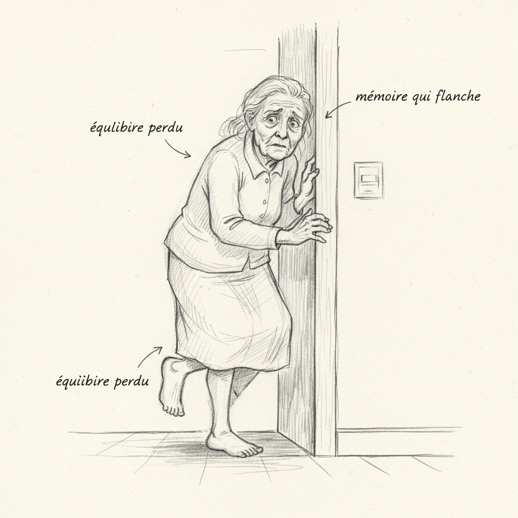
The second cause is malabsorption. Hypochlorhydria, meaning insufficient hydrochloric acid production by the stomach, is a major factor. And this point is crucial to understanding why supplementation so often fails: standard iron tablets (ferrous sulfate) simply can’t dissolve without gastric acid. Hypothyroidism itself slows gastric secretion[^8], creating a vicious circle between thyroid and iron absorption. Hypochlorhydria is extremely common in people over 40, in those taking PPIs long-term, and in those suffering from chronic stress. If you’ve been taking omeprazole for years, there’s a good chance your iron absorption is compromised. In elderly people, hypochlorhydria (insufficient acid production) or even achlorhydria (complete absence of acid) is so common that anemia symptoms (fatigue, pallor, cognitive decline) are often attributed to normal aging. How many exhausted grandparents are actually anemic without knowing it. Add that prolonged use of aspirin and nonsteroidal anti-inflammatory drugs (NSAIDs), very common in seniors, causes chronic digestive bleeding representing a loss of 50 to 60 mg of iron per month.
Intestinal dysbiosis is another cause of malabsorption. An unbalanced microbiota, chronic candidiasis, undiagnosed celiac disease, intestinal permeability: all of this disrupts nutrient absorption, including iron. When I see an anemic patient whose iron supplementation isn’t working, I systematically look at the intestines. That’s often where the key to the problem lies.

B12 and folate (B9) deficiency is the third major cause. Without B12 and B9, erythroblasts can’t multiply correctly. It’s megaloblastic anemia. Strict vegetarians are at high risk for B12 deficiency. Biermer disease, an autoimmune condition, is another classic cause. And certain medications (metformin, PPIs) decrease B12 absorption over time.
Chronic inflammation, discussed with hepcidin, is the fourth cause. Ferritin may be normal or even high, but functional iron is insufficient. Excessive losses are the fifth cause: heavy periods, occult digestive bleeding, intense exercise (effort hemolysis). Finally, rare genetic causes (thalassemia, sickle cell disease) fall under hematology care.
Do you have to eat meat to escape anemia?
Dietary iron exists in two forms. Heme iron, present in animal products (red meat, organ meats, blood sausage), is absorbed at a rate of 20 to 25 percent. Non-heme iron, present in plants (lentils, spirulina, pumpkin seeds), is absorbed at only 2 to 5 percent. The difference is considerable.
But this doesn’t mean vegetarians are doomed to anemia. Siegenberg’s study demonstrated that vitamin C spectacularly increases non-heme iron absorption[^4], by reducing it from its poorly absorbable ferric form to its directly assimilable ferrous form. 100 mg of vitamin C (a kiwi or half a bell pepper) multiply iron absorption from a meal by 4.14 according to Iron Disorders Institute data. A squeeze of lemon juice on your lentils can triple or quadruple absorption. Another underappreciated promoter: beta-carotene (apricots, carrots, sweet potatoes, spinach) significantly improves iron absorption and, remarkably, is capable of overcoming the inhibitory effect of phytates and tannins. And a gram of meat has a promoting effect on non-heme iron absorption equivalent to 1 mg of vitamin C, even in small amounts in a vegetarian meal.
Conversely, certain compounds inhibit iron absorption, and the figures are striking. Coffee and cocoa inhibit absorption by 60 to 90 percent[^5]. Phytates (soy, beans, lentils, whole grains) reduce it by 50 to 65 percent[^6]. Calcium, at doses of 300 to 600 mg (the equivalent of a glass of milk), inhibits both heme and non-heme iron absorption. Eggs contain phosvitin, a protein that reduces iron absorption by 28 percent per meal. Oxalates (spinach, beets, nuts, chocolate, tea) form insoluble complexes. The Iron Disorders Institute is indeed emphatic: iron from spinach is not easily absorbed, contrary to the popular myth inspired by Popeye. The tannins in black tea, coffee and berries, the polyphenols in apples and herbal teas, all participate in this locking mechanism. This is why I systematically advise against drinking tea or coffee during meals. Waiting at least two hours after the meal is a simple gesture that can completely change iron absorption in a deficient person.
Naturopathic solutions: rebuilding the terrain
“When the organism is in a swamp, it’s pointless to chase mosquitoes: drain the swamp.” Pierre Valentin Marchesseau
In naturopathy, we don’t treat anemia. We treat the terrain that allowed anemia to establish itself. And it always begins with diet. Liver, blood sausage, sardines, lentils, chickpeas, spirulina, pumpkin seeds, raw cacao: these are the pillars of an iron-rich diet. But intake is useless if absorption fails. The first reflex is to ensure the digestive system works correctly.
Digestive support is the foundation of my approach. For hypochlorhydria, bitter herbs are remarkable allies. Gentian, milk thistle, artichoke stimulate gastric and biliary secretion. Apple cider vinegar diluted in a little warm water before the meal is another simple gesture that promotes mineral absorption.
Cofactors are essential. Vitamin C, between 500 and 1000 mg per day, is the number one cofactor for iron absorption. Copper, whose role in iron export via hephaestin is now well-established. Vitamin A improves the mobilization of ferritin reserves. And B12 and folates are indispensable for erythropoiesis. Zinc is involved in hemoglobin production and overall immune support. A Japanese study cited by the Iron Disorders Institute demonstrated that supplementing zinc and iron simultaneously is more effective than either alone.
A point on iron forms. A standard 325 mg tablet of ferrous sulfate actually contains only 100 mg of elemental iron, the rest being the counter-ion. Ferrous sulfate remains most commonly prescribed, but it’s also the least well-tolerated: nausea, constipation, black stools. Iron gluconate, often present in liquid forms, is generally better absorbed with fewer side effects. Carbonyl iron is preferred by some doctors for its lower toxicity if accidentally overdosed in children. Heme iron (Proferrin) causes fewer gastrointestinal issues. And iron bisglycinate is the form I prescribe in consultations: better absorbed and better tolerated than ferrous sulfate, it doesn’t require gastric acid for assimilation, making it particularly suitable for people suffering from hypochlorhydria.
Golden rule: iron should be taken two hours before or after other medications. And beware: iron inhibits the effectiveness of thyroid medications, antibiotics, and certain antidepressants. If you’re on Levothyrox, space your iron dose by at least four hours. Recent research on hepcidin dynamics further suggests that dosing every other day might be more effective than daily dosing, because hepcidin rises after each iron dose and takes 24 to 48 hours to come back down. By spacing doses, you optimize each one.
In phytotherapy, nettle (Urtica dioica) is the quintessential plant for anemia. Rich in iron, vitamin C, chlorophyll, it provides both the mineral and the cofactors for its absorption. Spirulina is another pillar. Desmodium and milk thistle support the liver, the central organ of iron metabolism and hepcidin production. For inflammation management, curcumin, omega-3 (EPA/DHA) and quercetin can help lower hepcidin by reducing pro-inflammatory cytokines.
Tests that matter: beyond laboratory norms
The standard blood panel often limits itself to hemoglobin and ferritin. That’s insufficient. Ferritin tells you about reserves, but it’s also an acute phase reactant: it can be elevated in the presence of inflammation, infection, liver disease, even when iron stores are low. This is the classic inflammatory anemia trap. A complete blood count provides the MCV which allows classification of anemia: microcytic (low MCV, iron deficiency), macrocytic (elevated MCV, B12/B9 deficiency), or normocytic.
Transferrin saturation (saturation coefficient) is a marker I consider essential. The calculation is simple: fasting serum iron divided by total iron-binding capacity (TIBC), multiplied by 100. The normal range is between 25 and 35 percent. Below 20 percent, it’s iron deficiency or inflammatory anemia. Above 45 percent fasting, caution: you’re entering the iron overload zone. TIBC alone already provides valuable information: it’s elevated (above 400-450 mcg/dL) in iron deficiency because the body makes more transporters to capture the little iron available. It’s low in inflammatory anemia because iron is abundant but sequestered.
Reticulocytes indicate whether the bone marrow is responding correctly. The soluble transferrin receptor (sTFRC) is a marker of erythropoietic expansion and iron deficiency, independent of inflammation. The sTFRC/log ferritin ratio is the most reliable tool for diagnosing iron deficiency in an inflammatory context, an exam fewer than 5 percent of doctors order. And CRP tells you if you’re in inflammatory terrain, which changes the entire interpretation of your panel.
A point close to my heart: laboratory norms are not health norms. Doctor Hertoghe explained this very well. A ferritin of 15 ng/mL is “within normal range” for the lab, but it’s already symptomatic in clinical practice. The Iron Disorders Institute considers that a healthy adult should be between 25 and 75 ng/mL. In naturopathy, the optimal zone is between 50 and 100 ng/mL. It’s the difference between “not sick” and “in good health,” and that’s the entire philosophy of naturopathy.
Want to assess your iron deficiency risk? Take the iron deficiency questionnaire in 2 minutes. If you also suspect a thyroid problem, the Claeys test completes the panel.
The other side of the coin: when iron becomes poison
I can’t write a complete article on anemia without addressing iron overload. Because too much iron is as dangerous as too little. When transferrin saturation exceeds its capacity, non-transferrin-bound iron (NTBI) begins circulating freely in plasma. This free iron is a formidable pro-oxidant. It enters hepatocytes via the ZIP14 transporter, into cardiomyocytes by other pathways, and it generates reactive oxygen species that cause fibrosis, cirrhosis, heart failure and diabetes.
The Iron Disorders Institute alerts to an underappreciated fact: women become vulnerable to iron overload as soon as menstruation stops, whether through menopause, hysterectomy or long-term hormonal contraception. Hereditary hemochromatosis (HFE C282Y mutation, the most common in Caucasians) can remain silent for decades in menstruating women, then emerge abruptly after menopause. Too much iron can cause premature heart attacks, diabetes, liver disease, osteoarthritis, osteoporosis, and hormonal imbalances.
In elderly people, excess iron “can overwhelm the body’s natural storage capacity, leading to oxidative stress, tissue damage and accelerated aging” according to the Iron Disorders Institute. The paradox is that some patients present simultaneously with tissue iron excess AND anemia, because excess iron has damaged the kidneys or pituitary, reducing EPO production needed to make red blood cells. This is why a complete panel with transferrin saturation and ferritin is essential before any supplementation.
Giving your body the tools for its repair
Anemia is not inevitable. It’s a signal saying something isn’t working in the chain of iron absorption, transport, or recycling. A stomach not producing enough acid. An intestine no longer absorbing. Chronic inflammation locking up iron. Missing cofactors.
Naturopathy offers a holistic approach that doesn’t limit itself to iron supplementation. It works on digestive terrain, restores cofactors, manages inflammation, and supports the body’s elimination organs. The number of patients I’ve seen raise their ferritin after months of failed supplementation, simply by working on the stomach and intestines, is countless.
To support remineralization, a Hurom juice extractor allows you to prepare concentrated green juices in iron non-heme and cofactors (beet, spinach, parsley, lemon) with optimal assimilation thanks to cold pressing (-20% with the code francoisbenavente20). On the supplementation side, Sunday Natural offers iron bisglycinate and pharmaceutical-quality B12 (-10% with the code FRANCOIS10). Find all my partnerships with exclusive promo codes.
If you want personalized support, you can book a consultation.
To go further
- Iron: the silent deficiency that exhausts your body
- Low ferritin and hair loss: the link your doctor ignores
- Vitamin B12 (cobalamine): methylation, neurology and pernicious anemia
- Alzheimer’s: the metabolic disease you can prevent 20 years before
Want to assess your status? Take the free vitamin B12 deficiency questionnaire in 2 minutes


Laisser un commentaire
Sois le premier à commenter cet article.