His name is Marc, he’s 52 years old, and his cardiologist just prescribed him a statin because his LDL is at 1.65 g/L. “My cholesterol is too high, apparently.” When I ask him if he’s had a fatty acid profile test, he looks at me like I’m speaking Chinese. When I ask how many times a week he eats fatty fish, he thinks: “Smoked salmon on Sundays, sometimes. Is that enough?” His erythrocyte omega-3 index, which I requested, came back at 3.8%. His arachidonic acid/EPA ratio is at 14. His cell membranes are rigid as cardboard. And no one ever told him.
We talk a lot about omega-3s as “anti-inflammatory.” It’s true, but it’s reductive. Omega-3s are not anti-inflammatory medications. They are the architects of your cell membranes. And when the architects are absent, the whole building collapses: communication between cells, hormonal response, neurotransmission, resolution of inflammation, blood fluidity, insulin sensitivity. Everything begins with the membrane. If you recognize yourself in the signs I’m about to describe, take the omega-3 deficiency questionnaire I use in my practice.
“Man does not die, he kills himself. Every cell in his body is a miniature universe, and the membrane surrounding it is the frontier between life and death.” Dr Catherine Kousmine, Be Well in Your Plate Until 80 and Beyond (1980)
The causes of omega-3 deficiency
The plasma membrane is not a simple sac containing cytoplasm. It is a dynamic structure composed of a double layer of phospholipids in which proteins float (receptors, ion channels, transporters, enzymes), cholesterol and glycolipids1. The “fluid mosaic” model of Singer and Nicolson (1972) describes this membrane as a two-dimensional lipid ocean where proteins move laterally, cluster into lipid rafts, change conformation to transmit signals. Each phospholipid has a hydrophilic head and two hydrophobic tails. The tails are fatty acids. If the fatty acids are saturated (no double bonds), the tails are straight, parallel, tightly packed. The membrane is rigid. If the fatty acids are unsaturated (with double bonds in cis configuration), the tails have bends. The membrane is fluid2.

DHA (docosahexaenoic acid, C22:6 n-3) has six cis double bonds. Six bends. It is the most “twisted” fatty acid in nature. When incorporated into a membrane phospholipid, it creates considerable space around it. Proteins embedded in the membrane can move, change conformation, interact with their ligands. Synaptic vesicles can fuse with the presynaptic membrane to release neurotransmitters. Insulin receptors can cluster into functional groups. Membrane fluidity is not an abstract concept: it is the physical condition of all cellular communication.
The first cause of deficiency is insufficient dietary intake of preformed EPA and DHA. Omega-3s belong to the essential fatty acids: the body cannot manufacture them. Alpha-linolenic acid (ALA, C18:3 n-3), the plant leader, must be converted to EPA (C20:5 n-3) and then to DHA by the enzymes delta-6-desaturase and delta-5-desaturase3. But this conversion is a bottleneck: only 5 to 10% of ALA is converted to EPA, and less than 1% to DHA. In women of childbearing age, conversion is slightly better due to the effect of estrogens on delta-6-desaturase expression, which partly explains the relative cardiovascular protection of women before menopause. But it remains insufficient to cover real needs.
The second cause, and probably the most devastating, is the imbalance in the omega-6/omega-3 ratio. Anthropological studies show that our hunter-gatherer ancestors consumed a ratio close to 1 to 1. Traditional Mediterranean diet reaches a ratio of 4 to 1. Modern Western diet is at 15 to 1, sometimes 20 to 25 to 14. Three factors explain this drift. The industrialization of vegetable oils rich in omega-6 (sunflower, corn, soy, grapeseed) used massively in processed products, ready meals, collective catering, industrial bakery. Intensive farming that replaced pasture grass (rich in ALA) with soy and corn meal (rich in omega-6): an egg from a chicken fed flax seed contains 300 mg of omega-3, an egg from a battery hen contains 30 mg5. And the drastic reduction in consumption of wild fatty fish.

Omega-6 and omega-3 use the same desaturase enzymes. If your diet provides twenty times more omega-6 than omega-3, the enzymes are monopolized by omega-6. The conversion of ALA to EPA and then to DHA is crushed by enzymatic competition. The biochemical result is a permanent shift toward the pro-inflammatory pathway: arachidonic acid (AA, omega-6) accumulates in membranes, COX-2 and LOX-5 enzymes convert it to PGE2 (pain), thromboxane A2 (aggregation), leukotrienes B4 (inflammation)[^6]. The membrane AA/EPA ratio, which should be below 3, rises to 10, 15, sometimes 20 in patients who never eat fatty fish. Marc, with his ratio of 14, is in the French average. It’s not normal. It’s ordinary.
The third cause is deficiency in desaturase cofactors. Delta-6-desaturase, the limiting enzyme of the entire cascade, requires zinc, magnesium, vitamin B6 (P5P) and iron[^7]. The same cofactors that are lacking in modern diet. As I detail in the articles on zinc and the thyroid, these deficiencies are nearly universal. Without these cofactors, even adequate intake of plant ALA will never be converted to EPA and DHA.
The symptoms of deficiency
Omega-3 deficiency does not manifest as a single, obvious symptom. It sets in quietly, over months, sometimes years, and affects multiple systems simultaneously because membrane fluidity is the condition of all cellular communication.
The first area affected is the cardiovascular system. Insulin receptors, embedded in the membrane of muscle and liver cells, must move laterally, dimerize and change conformation when insulin binds. In a rigid membrane (too much saturated fat, not enough DHA), the receptors are physically “stuck.” The cell receives the signal but cannot respond[^8]. This is one of the overlooked causes of insulin resistance: the receptors are functional but immobilized in a membrane that’s too viscous. Ion channels (sodium, potassium, calcium) also depend on membrane fluidity. In a rigid membrane, conformational changes are slowed. Heart rhythm can become irregular. Epidemiological studies show a direct correlation between erythrocyte omega-3 index and risk of sudden cardiac death from arrhythmia: an index above 8% reduces risk by 90% compared to an index below 4%[^9]. The GISSI-Prevenzione study (11,324 post-infarction patients) showed a 45% reduction in sudden death with 1 g of EPA+DHA per day. Excessive platelet aggregation, endothelial dysfunction and elevated CRP complete the cardiovascular picture.
The second area is the nervous system. DHA represents 30 to 40% of the fatty acids in neuronal membranes and up to 60% in the outer segments of retinal photoreceptors. The brain and eye are the two organs richest in DHA in the human body. Synaptic vesicles fuse with the presynaptic membrane to release neurotransmitters (exocytosis), and this process requires optimal fluidity. A DHA deficit slows exocytosis of dopamine, serotonin and noradrenaline[^10]. Meta-analyses show that omega-3 supplementation (EPA dominant, 2 to 3 g per day) significantly improves depression scores, particularly in forms with an inflammatory component. Cognitive decline, memory problems, brain fog, attention disorders are all associated with a low omega-3 index. Dry eyes are another frequent sign, related to DHA’s role in retinal membranes and tear film quality.
The third area is low-grade chronic inflammation. Excess omega-6 does more than stiffen membranes. It fuels systemic low-grade inflammation (SLGI) recognized as the terrain common to all chronic diseases[^11]. Arachidonic acid is stored in phospholipids at the sn-2 position. When an inflammatory signal arrives, phospholipase A2 releases AA, which is converted by COX enzymes to PGE2 and by LOX enzymes to leukotrienes LTB4. EPA, when present in sufficient quantity, competes with AA for these same enzymes. The series 3 eicosanoids that result (PGE3, TXA3) have much lower inflammatory activity: PGE3 is a hundred times less pro-inflammatory than PGE2[^12]. In practice, deficiency manifests as diffuse joint pain, morning stiffness, dry and dull skin, recurrent allergies, chronic fatigue that doesn’t improve with rest, and vulnerability to repeated infections. These are symptoms many of my patients attribute to age when they actually reflect a correctable membrane imbalance.
The fourth area is hormonal communication. Receptors for thyroid hormones, cortisol, estrogens are all membrane proteins or intracellular proteins whose function depends on the fluidity of the lipid bilayer. A rigid membrane slows hormonal signal transduction. This is why omega-3 deficiency can mimic or worsen functional hypothyroidism, cortisol resistance, or estrogen-progesterone imbalance. The thyroid panel can be “normal” while target cells don’t properly receive the hormonal message.
The micronutrients essential to omega-3s
Omega-3s don’t work alone. Their metabolism, membrane incorporation and oxidation protection depend on a network of cofactors without which all supplementation will be partially ineffective.
Zinc is the first cofactor of desaturases. Delta-6-desaturase, the limiting enzyme for ALA conversion to EPA and then DHA, is a zinc-containing metalloenzyme. With zinc deficiency, even a generous intake of plant ALA remains “blocked” upstream of the cascade. Zinc also plays a role in membrane structural stability and antioxidant protection (cofactor of Cu/Zn-SOD superoxide dismutase). Supporting dosage: 15 to 30 mg of zinc bisglycinate per day[^13].
Magnesium is the second critical cofactor. It participates in the activity of delta-6-desaturase and delta-5-desaturase, in cell membrane stability and in over 300 enzymatic reactions. Magnesium deficiency, which affects more than 70% of French people according to the SU.VI.MAX study, is therefore a direct aggravating factor of functional omega-3 deficiency. Supporting dosage: 300 to 400 mg of magnesium bisglycinate per day.
Vitamin B6 in its active form pyridoxal-5-phosphate (P5P) is a cofactor for both desaturases. It is also essential for serotonin synthesis, which explains why omega-3 deficiency and serotonin deficit so often coexist in consultation. Oral contraceptives and PPIs (proton pump inhibitors) are two major destroyers of B6. Supporting dosage: 25 to 50 mg of P5P per day.
Iron is a cofactor of delta-6-desaturase. Low ferritin (below 50 ng/mL) compromises not only oxygen transport but also the conversion of plant omega-3s to EPA and DHA. This is a point I systematically check in patients who have fatigue, pallor and dry skin.
Vitamin E (mixed tocopherols) is the major lipophilic antioxidant of cell membranes. Polyunsaturated fatty acids, particularly DHA with its six double bonds, are extremely vulnerable to peroxidation by free radicals. Supplementing omega-3 without protecting membranes with vitamin E is like filling a leaky tank. Vitamin E inserts itself into the lipid bilayer and stops the chain reaction of peroxidation. Dosage: 200 to 400 IU of mixed tocopherols (alpha, beta, gamma, delta) per day[^14].
Selenium is a cofactor of glutathione peroxidase (GPx), the enzyme that neutralizes lipid hydroperoxides in membranes. Without selenium, peroxides accumulate and degrade membrane PUFAs. This is why populations with high consumption of fatty fish (Japan, Iceland) also have high selenium intakes via the same fish. Dosage: 100 to 200 micrograms of selenomethionine per day. And vitamin C, water-soluble, regenerates oxidized vitamin E at the membrane surface, completing the antioxidant trio of vitamin E / selenium / vitamin C.
Dietary sources
The dietary strategy rests on a dual movement: increasing omega-3 intake and simultaneously decreasing omega-6 intake.
Small fatty fish are the reference source: sardines, mackerel, herring, anchovies. Small because they accumulate less mercury and persistent organic pollutants (PCBs, dioxins) than large predators (tuna, swordfish, shark). A tin of sardines in olive oil provides approximately 1.5 g of EPA+DHA. Three servings of small fatty fish per week form the foundation of the dietary strategy. Wild salmon (not farmed on plant meal) is also an excellent source, with approximately 2 g of EPA+DHA per 150 g[^15]. Shellfish (oysters, mussels, shrimp) provide more modest amounts but contribute to overall balance while supplying zinc and selenium, two essential cofactors.
Plant-based ALA sources are complementary but do not replace marine sources. Rapeseed oil (omega-6/omega-3 ratio of 2 to 1) is the ideal table oil replacing sunflower. Camelina oil and flax oil are the richest in ALA (35% and 55% respectively), but recall that conversion to EPA and then DHA remains below 10% and 1%. Walnuts (2.5 g ALA per 30 g), chia seeds (5 g ALA per 30 g) and ground flax seeds (3.5 g ALA per 30 g) are useful daily additions. Flax must be ground or milled because whole seeds pass through the digestive tract without releasing their omega-3s.
Quality animal products also play an often underestimated role. An egg from a chicken fed flax seed (Bleu-Blanc-Coeur label in France) contains ten times more omega-3 than a standard egg. Beef raised on grass has an omega-6/omega-3 ratio of 2 to 1, compared to 20 to 1 for grain-fed beef. Butter from grass-fed cows, raw milk mountain cheeses are also underestimated sources of short-chain omega-3s and CLA (conjugated linoleic acid).
To reduce omega-6, you must eliminate sunflower, corn, soy and grapeseed oils in favor of olive oil (omega-9, neutral from an inflammatory standpoint) and rapeseed oil. Limit processed products, ready meals, industrial cookies, vegetable margarines. Read labels: “vegetable oil” in an industrial product almost always means sunflower or palm, therefore omega-6 or saturated fats.
The antagonists of omega-3s
Before adding omega-3s, it is essential to identify what destroys, blocks or wastes them. In naturopathy, Marchesseau’s rule applies: first do no harm, meaning eliminate the causes before providing solutions.
Trans fatty acids are the most dangerous antagonists. Derived from partial hydrogenation of vegetable oils (industrial margarines, pastries, cookies, fast-food), trans slip into cell membranes in place of omega-3s and stiffen them without any of the functional properties of cis fatty acids. Worse, they directly inhibit both desaturases, blocking the conversion of ALA to EPA and DHA[^16]. A single fast-food meal can provide 5 to 10 g of trans. The European Union has limited industrial trans fats to 2 g per 100 g of fat since 2021, but old formulations are still circulating and frying remains a major source.
Excess omega-6 is the second antagonist, through direct enzymatic competition. Omega-6 and omega-3 use the same desaturases and elongases. When diet provides twenty times more omega-6, the enzymes are saturated by the linoleic acid to arachidonic acid pathway, and the omega-3 pathway is crushed. Correcting the ratio is therefore at least as important as supplementing with EPA/DHA.
Alcohol directly inhibits delta-6-desaturase. Regular consumption, even “moderate” (two glasses of wine per day), significantly reduces the capacity to convert ALA and promotes peroxidation of membrane PUFAs. Alcohol is a dual antagonist: it blocks synthesis and accelerates destruction. Tobacco acts by the same mechanism of enzymatic inhibition, aggravated by the massive generation of free radicals that oxidize membrane PUFAs.
Oil refining destroys heat-sensitive omega-3s. Rapeseed or flax oil cold-pressed retains its omega-3s intact. The same oil refined (heated to 200-240 degrees, deodorized, decolorized) has lost nearly all of its omega-3s and contains traces of trans. High-temperature cooking (frying, grilling, high heat pan) also destroys omega-3s in food. Grilled fish loses 50 to 80% of its EPA/DHA. Gentle cooking is the only method that preserves polyunsaturated fatty acids.
Refined sugar and insulin resistance form a vicious circle with omega-3 deficiency. Chronic hyperinsulinism inhibits desaturases. Rigid membranes worsen insulin resistance (receptors don’t move). Insulin resistance further inhibits desaturases. Type 2 diabetes is both a consequence of and an aggravating factor for omega-3 deficiency.
Statins, paradoxically, reduce the synthesis of coenzyme Q10 (which protects membranes against oxidation) and do not correct the membrane AA/EPA imbalance. Marc, who was being prescribed a statin for his LDL, had a membrane problem, not a cholesterol problem. Non-steroidal anti-inflammatory drugs (NSAIDs) block COX, treating the inflammatory consequence without correcting the membrane cause.
The forgotten causes of deficiency
Beyond dietary deficit and antagonists, certain causes of omega-3 deficiency escape the radar of conventional medicine.
Aging progressively reduces desaturase activity. After age 50, the conversion of ALA to EPA and DHA, already weak in young subjects, decreases further by 30 to 50%[^17]. This is why the need for preformed EPA and DHA increases with age, at the very time when fish consumption often decreases (chewing problems, fear of mercury, fixed eating habits). Dr Thierry Hertoghe emphasizes the link between membrane aging and hormonal decline: hormonal receptors lose their mobility in membranes depleted of DHA, which mimics a hormonal deficit even when circulating hormones are within normal ranges.
Erythrocyte omega-3 index is the test no one prescribes but everyone should get. Red blood cells have a lifespan of 120 days, and their membrane composition reflects intake from the past four months, making it a far more reliable marker than plasma levels that fluctuate meal to meal[^18]. An index below 4% is associated with high cardiovascular risk. Between 4 and 8%, risk is intermediate. Above 8%, protection is optimal. The French average is 4.5%. The Japanese average is 9.5% (Japan has one of the lowest rates of cardiovascular disease and depression in the world). The membrane AA/EPA ratio completes the picture: the goal is below 3, above 5 inflammation is significant, above 10 it’s major. The complete profile (available from Barbier, Synlab, 80 to 120 euros not reimbursed) also measures the percentage of saturated, monounsaturated, omega-6 and omega-3 fatty acids, and the membrane fluidity index[^19].
Insulin resistance and metabolic syndrome block desaturases through a direct inhibition mechanism linked to chronic hyperinsulinism. The obese, diabetic or prediabetic patient therefore combines increased omega-3 need (chronic inflammation, membrane rigidity) and increased inability to synthesize them from plant precursors. This is a metabolic trap requiring preformed EPA and DHA intake.
Chronic stress and permanent cortisol elevation accelerate membrane fatty acid turnover and increase omega-3 consumption. Patients in burnout, adrenal exhaustion, or under intense work stress often present with a collapsed omega-3 index, even with adequate diet. Stress is an omega-3 burner.
Intestinal dysbiosis and intestinal permeability impair the absorption of fatty acids and cofactors (zinc, magnesium, iron, B6). An inflamed intestine absorbs dietary omega-3s poorly and converts plant ALA even more poorly. This is why I always start by restoring the intestinal barrier before hoping to durably correct the fatty acid profile.
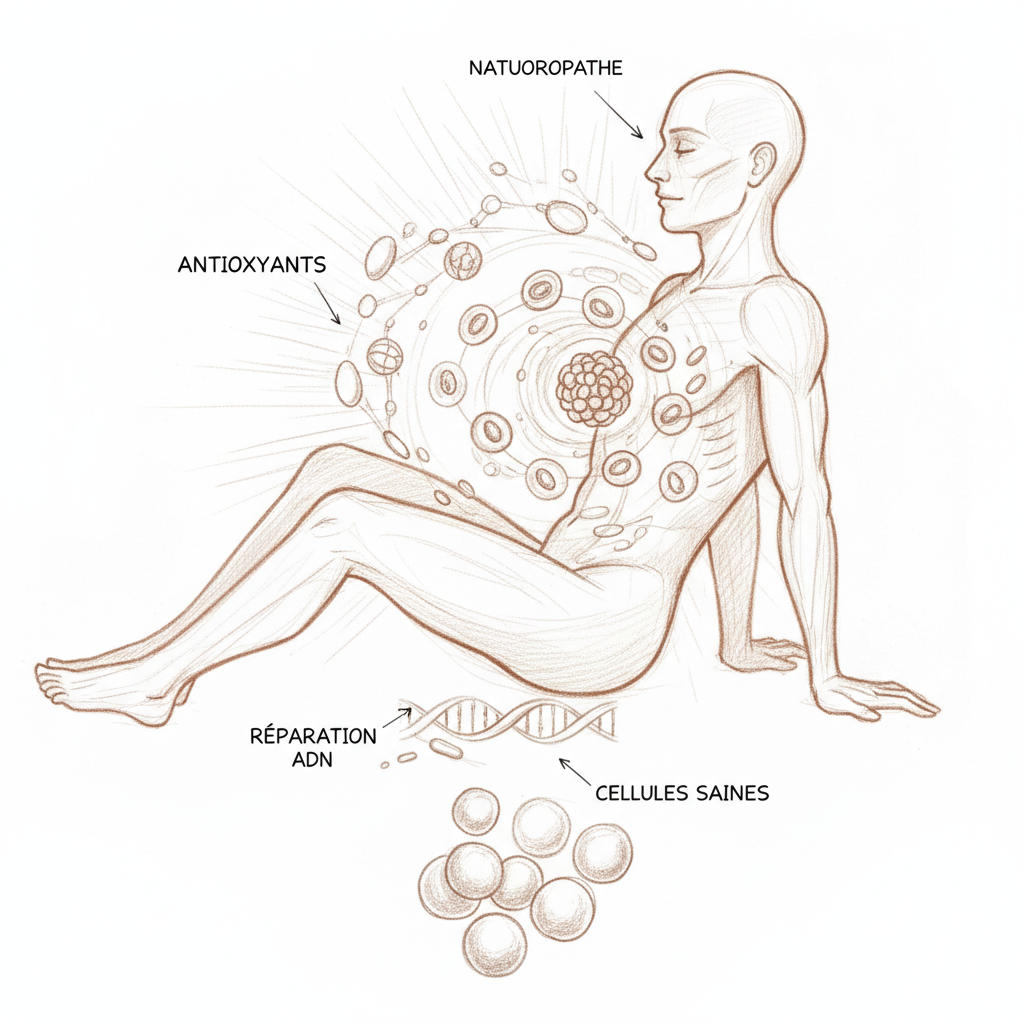
The most important discovery in lipidology over the past twenty years illuminates an additional forgotten cause: deficiency in active resolution of inflammation. For a long time, it was believed that inflammation simply died down. The work of Charles Serhan (Harvard Medical School) showed that there are specialized lipid mediators that actively extinguish inflammation and orchestrate tissue repair[^20]. Resolvins series E (derived from EPA) and series D (derived from DHA) inhibit neutrophil migration, stimulate phagocytosis of cellular debris and decrease pro-inflammatory cytokines. Neuroprotectins D1 (derived from DHA) directly protect neurons from apoptosis. Maresins stimulate tissue regeneration. Without sufficient EPA and DHA in membranes, the body cannot manufacture these mediators. Inflammation doesn’t go out. It becomes chronic. This is the vicious circle of SLGI: inflammation damages membranes, which lose their DHA, which reduces resolution production, which prevents inflammation from resolving, which damages membranes further.
Food supplements
When the fatty acid profile confirms a deficit (omega-3 index below 8%, AA/EPA ratio above 3), supplementation becomes necessary because dietary correction alone takes six to twelve months to normalize membranes.
Fish oil (EPA + DHA combined) remains the reference. The effective dosage is 2 to 3 g of EPA+DHA per day, not 2 to 3 g of fish oil (a 1 g fish oil capsule often contains only 300 mg of EPA+DHA, the rest being other fatty acids). You must therefore read the composition, not the capsule weight. The natural triglyceride form (TG) offers better bioavailability than the ethyl ester form (EE): intestinal absorption is 50 to 70% higher[^21]. Look for “TG form” or “triglyceride” on the label.
The EPA/DHA ratio should be adapted to the clinical situation. Dr Merran recommends an EPA/DHA ratio of 2 to 1 for inflammatory situations (joint pain, SLGI, depression, autoimmune diseases). EPA is the direct precursor of series E resolvins and anti-inflammatory series 3 eicosanoids. A 1 to 2 ratio (DHA dominant) is preferable for neurological and cognitive situations (cognitive decline, pregnancy, child brain development, dry eyes).
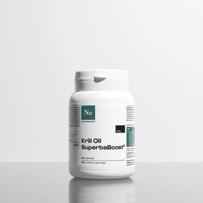
Krill oil is an interesting alternative. Krill omega-3s are in phospholipid form, which facilitates their direct incorporation into cell membranes (membranes are themselves made of phospholipids). Krill also provides astaxanthin, a powerful antioxidant that protects omega-3s from oxidation. Disadvantage: krill is less concentrated in EPA/DHA than fish oil, and more costly at equivalent dosage.
Algae omega-3s (Schizochytrium or Ulkenia microalgae oil) are the only vegan source of preformed EPA and especially DHA. This is the logical choice for vegetarians and vegans, and also for people who don’t tolerate fish oil (reflux, belching). DHA concentration is generally good (200 to 400 mg per capsule), but EPA content is often lower. Some brands now offer formulas enriched in algae EPA.
Duration of supplementation is something I systematically clarify in consultation. Red blood cell membranes renew in 120 days. Neuronal membranes renew more slowly. Expect three to six months of regular supplementation to observe normalization of the omega-3 index and significant clinical improvement. Marc took six months to go from 3.8% to 7.2%. It’s not a one-time treatment, it’s a terrain correction requiring consistency.
Antioxidant protection is inseparable from omega-3 supplementation. PUFAs are the fatty acids most vulnerable to peroxidation. Supplementing omega-3 without protecting membranes with antioxidants is to accelerate membrane oxidative stress. The minimal trio is: vitamin E (mixed tocopherols, 200 to 400 IU per day), selenium (100 to 200 micrograms per day, cofactor of glutathione peroxidase) and vitamin C (500 mg per day, which regenerates oxidized vitamin E)[^22]. Omega-3s should be kept refrigerated, away from light and air.
Marc’s complete protocol illustrates the naturopathic approach. Sardines or mackerel three times a week. Olive oil and rapeseed oil replacing sunflower. Supplementation with EPA/DHA 3 g per day (triglyceride form). Vitamin E 400 IU. Selenium 200 micrograms. Zinc bisglycinate 25 mg. Magnesium bisglycinate 400 mg. Elimination of all hidden sources of omega-6. At six months, his omega-3 index had risen to 7.2%. His AA/EPA ratio had gone from 14 to 4.5. His CRP had dropped from 3.2 to 0.8 mg/L. His LDL hadn’t moved, but his oxidized LDL had decreased by 40%. His fasting glucose had come back down from 1.05 to 0.92 g/L. He no longer had joint pain. And he slept better.
Marc didn’t take a statin. His cardiologist, seeing the results of the inflammatory panel and fatty acid profile, agreed to postpone the prescription. It’s not always the case. But that’s what should happen when you look at membranes before looking at cholesterol.
Your cells are islands. The membrane is the ocean that connects them. If the ocean is frozen, the islands no longer communicate. Omega-3s are not just another food supplement. They are the guardians of your fluidity. And your fluidity is your life.
If you’d like to take stock of your situation, you can schedule a consultation. For omega-3 supplementation, vitamin E, selenium and zinc, Sunday Natural offers pharmaceutical-grade formulas (code FRANCOIS10 for -10%). Find all my partnerships with exclusive promo codes.
To go further
- Cancer and nutrition: what micronutrition changes in the equation
- Cholesterol and cardiovascular disease: the real culprits your cardiologist isn’t looking for
- Alzheimer: the metabolic disease you can prevent 20 years before
- Anemia: understanding the deep causes and acting naturally
Sources
- Curtay, Jean-Paul. Nutriotherapy: Scientific bases and medical practice. 6th ed. Marco Pietteur, 2016.
- Mouton, Georges. Digestive Ecology: optimizing your intestinal ecosystem. Marco Pietteur, 2004.
- Kousmine, Catherine. Be Well in Your Plate Until 80 and Beyond. Sand/Tchou, 1980.
- Merran, Sarah. Fatty Acids and Membrane Fluidity. Diploma in Micronutrition, Nutrition, Prevention and Health (MAPS).
- Hertoghe, Thierry. The Hormone Handbook. 2nd ed. Luxembourg: International Medical Books, 2012.
- Masson, Robert. Dietetics of Experience. Guy Trédaniel, 2014.
“Every human being is rebuilt with food, baths and exercise. The rest is utopia.” Pierre-Valentin Marchesseau
Scientific References
Footnotes
-
Singer SJ, Nicolson GL. “The Fluid Mosaic Model of the Structure of Cell Membranes.” Science 175, no. 4023 (1972): 720-731. PMID: 4333397. ↩
-
Merran S. Fatty Acids and Membrane Fluidity. Diploma in Micronutrition (MAPS). Slide 24: “Nature of fatty acids in phospholipids: SFA → straight tails → rigid membrane. PUFA → bends (cis) → fluid membrane. DHA (C22:6 n-3): 6 double bonds, maximum fluidity.” ↩
-
Burdge GC, Calder PC. “Conversion of Alpha-Linolenic Acid to Longer-Chain Polyunsaturated Fatty Acids in Human Adults.” Reprod Nutr Dev 45, no. 5 (2005): 581-597. PMID: 16188209. ↩
-
Simopoulos AP. “The Importance of the Omega-6/Omega-3 Fatty Acid Ratio in Cardiovascular Disease and Other Chronic Diseases.” Exp Biol Med 233, no. 6 (2008): 674-688. PMID: 18408140. ↩
-
Merran S. ↩


Laisser un commentaire
Sois le premier à commenter cet article.